Real-Time Imaging of Previously Unseen Protein Structures (No. 0201)
|
|
|
<< Back to all technologies |
Summary
A novel electron microscope approach that provides direct and real-time images of previously unseen micro sized protein structures.
The global protein imaging market size is projected to reach USD 1.7 billion by 2025, at a CAGR of 8.9%. The growth in this market is attributed to the increasing adoption of protein therapeutics, technological advancements in protein imaging instruments and consumables, and the increasing focus on miniaturization. However, current instruments are still unable to clearly image biologically important micro-sized membrane proteins. Here we present a promising imaging system developed by a group of researchers led by Prof. Tsumoru Shintake. The imaging system is based on a transmission electron microscope that uses a spiral electron beam sweep on a stationary protein molecule. As a result of the electron sweep and detection, it is possible to visualize, in real-time, a micro sized structure.
Applications
- Protein Imaging
- Drug Discovery
Advantages
- Micro protein particle imaging
- Real-time imaging
- Direct Analysis
Technology
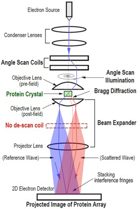
The technology is based on a system and method for obtaining a high-resolution image of the molecule array in a protein crystal by improving on techniques of Micro-ED, then translate the phase in Bragg diffraction to a structural phase, followed by magnifying the interference fringe through a beam expander, and understanding of the image output based on Ewald sphere and lattice point. To realize the direct imaging of a micro crystal, the core of this novel apparatus includes an electron source, angle scan coils, a beam expander and a detector. In this novel arrangement, no de-scan coil is required for beam expansion.
Media Coverage and Presentations
CONTACT FOR MORE INFORMATION
![]() Graham Garner
Graham Garner
Technology Licensing Section
![]() tls@oist.jp
tls@oist.jp
![]() +81(0)98-966-8937
+81(0)98-966-8937


