

FY2015 Annual Report
Quantum Wave Microscopy Unit
Professor Tsumoru Shintake
Abstract
1. R&D on diffraction electron microscope which uses the quantum wave interference associated with low energy electron as well as the technique of electron holography. We are developing an electron microscope capable of damage-free imaging to viral samples in sub-nanometer resolution with the use of a few tens of keV electron beam and the phase-retrieval algorithms. Prior to 2015 the unit developed an electron microscope, DMF4000, that covers the energy range in few hundred eV to 30keV, in collaboration with HITACHI High Tech Science, and the electron beam transfer system to image the diffraction pattern. In 2015 we have adopted in-line holography method and succeeded in reconstructing gold particles in high resolution. We have also investigated properties of graphene sheet with environmental TEM, and cleaning methods of its surface in order to use graphene film as the support of the viral sample.
2. We proposed a novel ocean-current turbine as a sustainable energy source in 2012. The turbine will convert the kinematic energy of an ocean-current into electricity. Thus far, in order to verify our ocean-current turbine design, our group conducted various towing tests and mooring experiments by scale models. As the next step, we planed developing a 10 kW model to perform a feasibility study with tidal current. In FY 2015, we conducted two R&D items. First, we installed an observation floating body to observe tidal current and the floating body motion. Secondly, we designed a 10-kW ocean-current turbine. The basic plan of the turbine was created.
1. Staff
- Dr. Katsutoshi Shirasawa, Staff Scientist
- Dr. Cathal Cassidy, Staff Scientist
- Dr. Teruhisa Hirai, Staff Scientist
- Dr. Hidehito Adaniya, Postdoctoral Scholar
- Dr. Masao Yamashita, Postdoctoral Scholar
- Dr. Martin Philip Cheung, Postdoctoral Scholar
- Hideki Takebe, Technician
- Jun Fujita, Technician
- Junichiro Minami, Technician
- Chola Kalale, OIST PhD Student
- Ankur Dhar, OIST PhD Student
- Hitomi Taba, Research Administrator
- Ayumi Shimojima, Research Administrator
2. Collaborations
2.1 Theme: Development of the ocean-current turbine
- Type of collaboration: Joint research
- Researchers:
- Professor, Dr. Hidetsugu Iwashita, Hiroshima University
2.2 Theme: Development of novel specimen grids for Transmission Electron Microscopy (TEM)
- Type of collaboration: Joint research
- Researchers:
- Professor, Dr. Jun Mizuno, Waseda University
- Students, Hiroyuki Kuwae, Xu Bingyang, Waseda University
3. Activities and Findings
3.1 Development of the imaging method for electron diffraction microscope
In principle, by applying the method of iterative phase retrieval on coherent diffraction data, one can determine the phase of the diffraction beam, and reconstruct the real image of the sample object. This process works correctly if the object has a two dimensional structure. The phase of the diffraction beam from an object of three dimensional structure, however, cannot be determined uniquely. In conventional bio-sample imaging with electron microscope, sample damage due to the impact by the impinging beam is an issue. One would stain the target sample for protection, however, it will limit the attainable resolution due to the size of staining particles in few nm. Ice-embedding the sample is also used widely for conventional TEM, however, this requires millions of images to be averaged over to obtain high resolution image, and the averaging method would fail if the bio-sample has asymmetric structure. To circumvent these issues, we have been developing a low energy diffraction microscope, DMF4000, to image bio-sample in a single-particle level. This is capable of SEM imaging and FIB etching, and the electron beam transport system with a 8k8k CMOS detector to obtain the diffraction patterns of scattered electron of low energy beam, smaller than a few keV (Fig.1).
We have adopted in-line holography in Fresnel mode in the range of 10 to 20keV. This method allows for the amplitude and phase part of the wave function retrieved at the same time with a single back propagation and few iterations of phase retrieval. Far simpler and more stable than conventional image reconstruction methods used in far field electron diffraction. With this method we have obtained the resolution about 1nm in reconstructed image of gold nano-particles. And it is expected that the phase change of the wave function of low energy beam caused by the bio-sample may be more pronounced compared to the conventional high energy TEM. The real and imaginary parts of the wave function would complementary contribute the high resolution imaging, and this would enable single-particle imaging to viral sample without averaging (Fig.2).
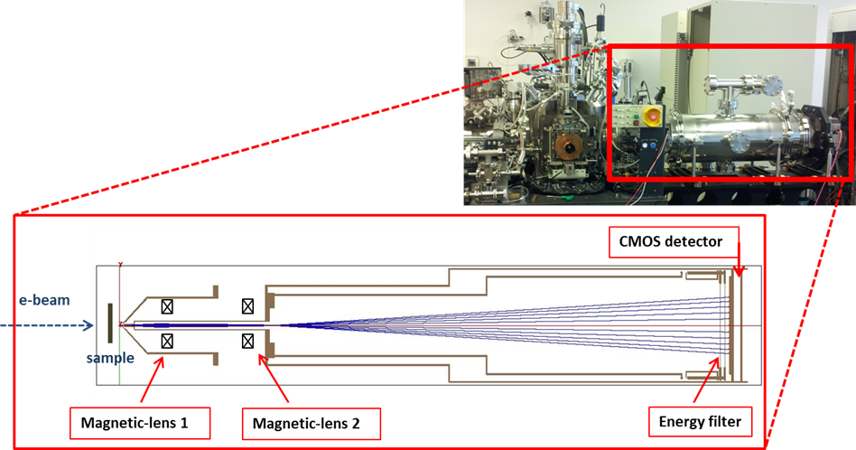
Figure 1. Top: Low energy diffraction electron microscope; DMF4000. Bottom: electron trajectory simulation for in-line holography
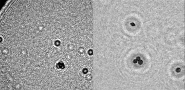
Figure 2. Left: holography image of gold particles on carbon support film, Right: reconstructed image of left
Free-standing graphene as a viral sample support film
Our unit has been focusing on low voltage holographic imaging of nanoparticle specimens on an ultraclean graphene support film. Graphene is only single atom thick and is electrically conductive; therefore it can be an excellent specimen support for the low voltage transmission imaging of nanometer-sized objects such as virus particles. We have optimized the fabrication of an ultraclean graphene support film on electron microscope grids and also devised an efficient approach to enriching hydrophilic specimen particles on a hydrophobic graphene film. Fig. 3 shows the scanning transmission electron microscopy (STEM) images of unstained bacteriophage T4 recorded at 20 kV on the graphene (left) and conventional carbon (right) support films. The improvement in the image signal-to-noise by the use of the graphene support film is clearly appreciable in the comparison of these images.
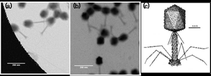
Figure 3. 20 kV STEM images of unstained bacteriophage T4 on ultraclean graphene (a) and amorphous carbon (b). (c) The structure of bacteriophage T4 (Leiman PG et al., CMLS Cell Mol. Life Sci. 60, 2356-2370. (2003)).
Free-standing graphene transfer, cleaning, virus-encapsulation
In order to achieve high-resolution images of biological specimens in transmission electron microscopy (TEM), two key aspects are of paramount-importance; image contrast and sample preservation. These facets become increasingly important as electron energy decreases and become limiting factors for low-energy electron microscopy. In traditional cryo-TEM, biological samples are embedded in ice to abate the negative impacts of sample dehydration and electron beam damage. Image contrast is a function of ice thickness and stringent control of this parameter greatly affects resolution. However, achieving thin ice can be extremely challenging and time consuming. As an alternative to traditional ice embedment, we have been investigating the concept of graphene cell encapsulation. In this work, we have developed a unique method to produce ultra-clean monolayer graphene TEM grids, producing a near-transparent support film (Fig.4).
Furthermore, we have developed a method to encapsulate biological samples within a thin graphene cell, maintaining the sample in a hydrated environment, while at the same time causing little impact to electron transmission. These advances have allowed us to produce high-contrast images of hydrated tobacco mosaic virus (TMV) with electron energies as low as 10 keV (Fig. 5).
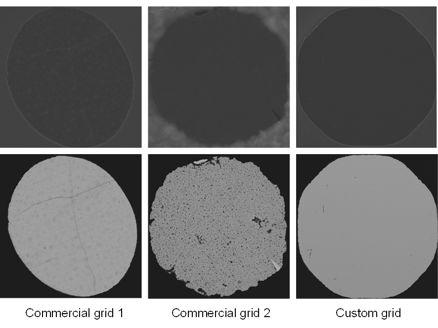
Figure 4. SEM (top) and STEM (bottom) comparisons of our custom monolayer graphene grids to commercially available graphene grids. Custom grids were produced by a PMMA-free direct transfer approach. All images recorded at 20 KeV.
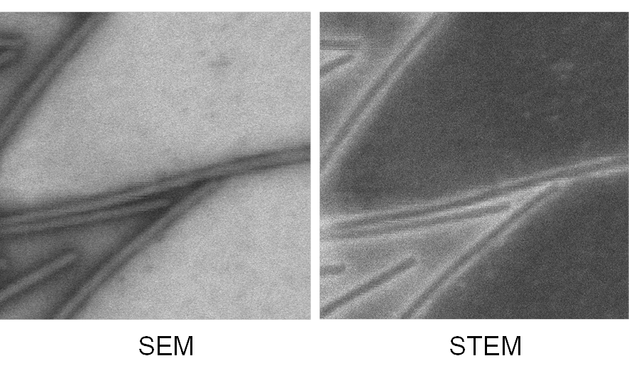
Figure 5. SEM and STEM images of TMV encapsulated between monolayer graphene sheets. Images recorded at 10 KeV
In-situ graphene characterization with environmental TEM
Synthesis and characterization of graphene has seen enormous research activity and significant progress in recent years [1]. However, for application of graphene as a transparent TEM grid, reproducible synthesis of large area free-standing single layer graphene, free of residual organic contamination, remains challenging [2]. In this study, we conducted some preliminary investigations on the effect of in situ TEM annealing on free-standing single layer graphene sheets, and establish some working procedures, target parameters and boundary conditions for base vacuum quality, electron beam exposure history, annealing temperature and ambient gas (H2/O2) atmosphere (Fig.6).
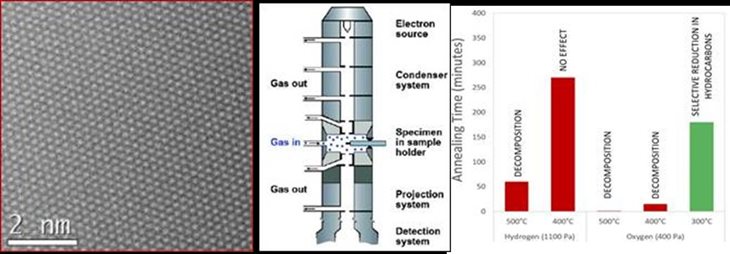
Figure 6. Left: pristine single layer graphene, Middle: schematics of in-situ TEM experiment, Right: Results of annealing with respect to gas species, annealing temperature and time
[1] Ferrara et al., Nanoscale, 2015 (7) 4598.
[2] Tyler et al., J. Phys. Chem. C, 2015, 119 (31), pp 17836–17841
3.2 Ocean Power Generation Group
Observation floating body
Our ocean-current turbine will work in a middle layer of an ocean current to avoid surface waves. In FY 2014,in order to obtain real tidal current data, we installed the observation floating body into the northern sea of Okinawa. The floating body is equipped with depth meters, thermometers and accelerometers. On the sea surface, the communication buoy is working to send the measured data to our database. In addition, the buoy has an ADCP (Acoustic Doppler Current Profiler) to measure current speed profile. In FY 2015, we conducted the observation and obtained the one-year data. We use the measured data to optimize our turbine performance and mooring system. Fig. 1 shows the current velocity and distribution measured by the ADCP.

Figure 1. Current velocity and distribution measured by the ADCP.
Basic design of 10-kW turbine
To propose a feasibility study, we designed a 10 kW ocean-current turbine. The design parameters are listed in TABLE I. All of the power-generating components, i.e., the electric generator, gear box and drive train are placed in the nacelle. Fig. 2 shows a design drawing of the nacelle. The nacelle head is the mooring point. The nacelle structure supports the load of the turbine’s thrust force. In addition, the float and counterweight are connected to this nacelle. Therefore, the nacelle is required to withstand the rotor torque.
Ideally, the electric generator should be driven directly by the turbine because it helps in avoiding transmission loss, maintenance work, and mechanical failure. However, turbine rotation is slow in water; therefore, a planetary gear is installed to increase the rotation speed of the generator. To keep the nacelle watertight, a mechanical seal system is installed on the rotor axis. Fig. 3 shows a 3D diagram of the turbine.
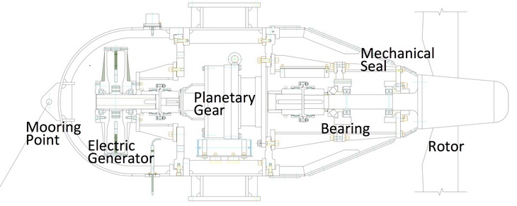
Figure 2. Design drawing of 10-kW turbine nacelle in a cross-sectional view.
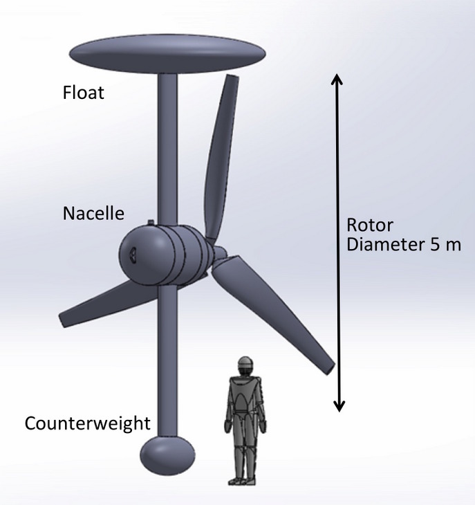
Figure 3. 3D diagram of 10-kW ocean-current turbine.
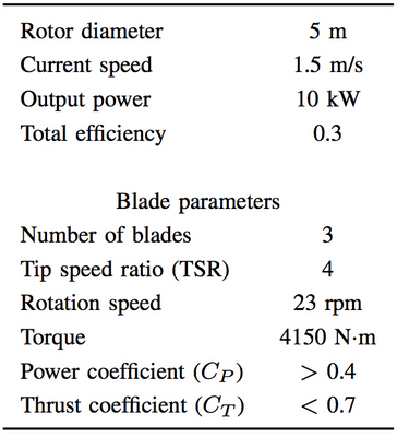
Table 1. Design parameters of 10-kW ocean-current turbine.
4. Publications
4.1 Journals
Nothing to report
4.2 Books and other one-time publications
Nothing to report
4.3 Oral and Poster Presentations
- Shintake, T., Japan, country of water, generates energy from the ocean, in 9th Green International Linear Collider Accelerator Working Group, Tokyo (2015).
- Shintake, T., Ocean Energy Research at OIST, in 2015 International Symposium on Energy Technology and Strategy, Taiwan, Cheng King Univ (2015).
- Shintake, T., Japan, country of water, generates energy from the ocean, in Surface Interface Spectroscopy 2015, Saitama prefecture (2015).
- Shirasawa, K., Minami, J., Iwashita, H., Shintake, T., Performance test of an ocean-current turbine at a circulating water channel, in 37th Japan Wind Energy Association, Tokyo (2015).
- Shirasawa, K., Development of an ocean-current turbine -Collaborative research with Hiroshima University, Hiroshima University (2015).
- Shirasawa, K., Minami, J., Iwashita, H., Shintake, T., Development of an ocean-current turbine at OIST, in International Research Institute for Applied Mechanics Symposium of Kyushu Univ on Ocean Renewable Energy Technologies and Related Fluid Dynamics Research, Kyushu University, Fukuoka Prefecture (2015).
5. Intellectual Property Rights and Other Specific Achievements
Nothing to report
6. Meetings and Events
Nothing to report
7. Other
Nothing to report.