

FY2021 Annual Report
Quantum Wave Microscopy Unit
Professor Tsumoru Shintake
Abstract
Microscopy group
Our collaboration with Dr. Latychevskaia in PSI yielded a publication in Ultramicroscopy on Bragg holography of nanocrystals. During this year, Dr. Cassidy also provided dedicated support to the wider OIST community from Sept-Nov, providing training and advice for multiple users from Narita, Qi, Wolf and Dani units in utilization of the Environmental TEM. We also collaborated with Prof. Nakagawa from University of the Ryukyus to perform in-situ measurements of hydrogen storage materials. We submitted a publication (currently under review) on mean free paths in electron holography, as conclusion of Dr. Cassidy’s Kakenhi project which finishes this year. We concluded a Collaborative Research Agreement (CRA) with local company, Acrorad, to continue the CdTe holography research theme. Additionally, we submitted a manuscript related to the thesis work of Dr. Ankur Dhar (our former PhD student), on electron holography of magnetic monopoles.
New working principle of the electron microscope was proposed by Dr. Shintake in 2021 (https://arxiv.org/abs/2105.03850), which will make possible to directly image molecule array inside the protein crystals. Dr. M. Yamashita and Dr. H. Adaniya studied sample preparation method suitable for this microscopy. JEOL Ltd. Japan started R&D on this microscope independently at their laboratory.
New telecentric optics for EUV lithography was proposed by Dr. Shintake, in January 2022 (https://arxiv.org/abs/2201.06749), which will solve the fundamental problem (mask 3D effect) in the current EUV photolithography. If it works, 5 nm or even narrower line process will be realized in the semiconductor device fabrication in future. It has been filed as US provisional patent.
Ethanol project group
At the beginning of current COVID-19 pandemic, Shintake proposed possible infection prevention and cure via ethanol inhalation in March 2020 (https://arxiv.org/abs/2003.12444). Under collaboration with Ishikawa Unit, we study disinfection mechanism due to ethanol (EtOH) in vitro, and trials on the infected mice with influenza to see recovering phenomena by ethanol vapor inhalation. We have very important results as follows: EtOH concentration required to disinfect the influenza virus is 20 %v/v at body temperature of 37 deg.C, which is much lower that the well-known value of 30%v/v at room temperature. Evidence of efficacy on treatment has been obtained, summary report will be published.
OIST Wave energy converter group
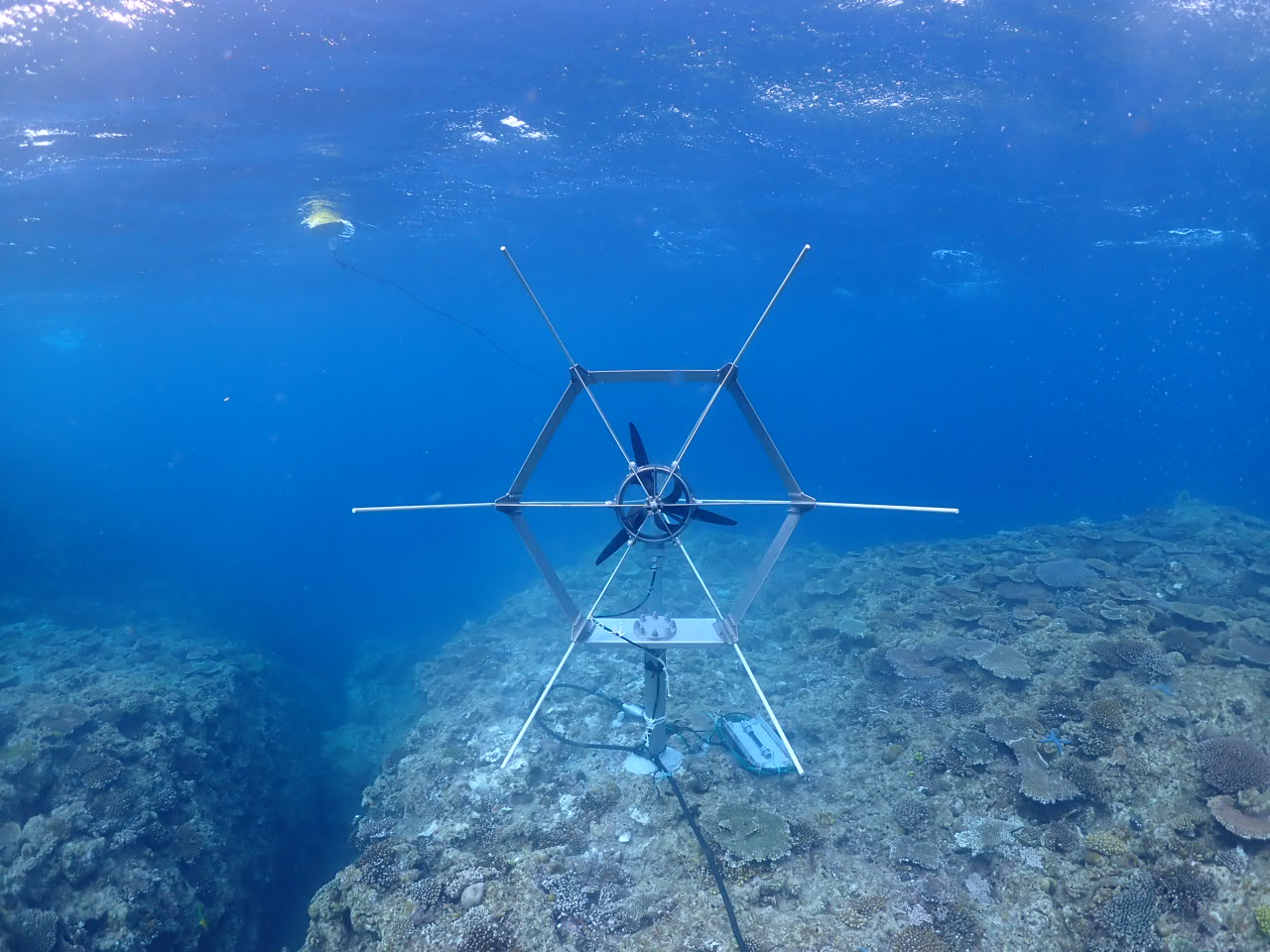
New concept of wave energy device was proposed by Dr. Shintake in 2021 (September 30, 2021, US Provisional 2021-161724, Surface Ocean Wave Turbine) We have performed four times of ocean experiments on turbine, towing by boat at Maeda area. YAMAHA MOTOR CO.,LTD. jointed to our project, donated four HARMO (electric outboard motor) to OIST. We assembled one HARMO with three-blade turbine, and installed in seabed at 3 m deep, near drop-off in front of OIST Marine Science Station at Seragaki in April 2022.
1. Staff
- Dr. Cathal Cassidy, Staff Scientist
- Dr. Masao Yamashita, Staff Scientist
- Dr. Ryusuke Kuwahara, Staff Scientist
- Dr. Yuichiro Hamate, PosDoc
- Dr. Krisna Pawitan, PosDoc
- Dr. Hidetoshi Adaniya, Research Unit Technician
- Dr. Martin Philip Cheung, Research Unit Technician
- Seita Taba, Research Unit Technician
- Jun Fujita, Research Unit Technician
- Shuji Misumi, Research Unit Technician
- Hideki Takebe, Specialist
2. Collaborations
2.1 Comparison of low energy microscopy techniques
- Description : At OIST, we have developed specific competence in preparing low-kV compatible ice-embedded samples. We are collaborating with Hitachi to understand how such samples appear in high-end commercial microscopes, and how the performance compares with low-energy holographic as well as STEM imaging of our developed microscope. Type of collaboration: Joint research
- Researchers :
- HITACHI
- Dr. Hidetoshi Adaniya, OIST
- Dr. Martin Philip Cheung, OIST
2.2 Imaging magnetic monopoles in spin ice
- Type of collaboration: Joint research
- Researchers:
- Dr. Ankur Dhar, SLAC
- Dr. Ludovic Jaubert, CNRS Bordeaux
- Prof. Nic Shannon, OIST
- Dr. Cathal Cassidy, OIST
2.3 Electron holography & diffraction for nanocrystal structure determination
- Description: At OIST, we are exploring a hybrid Bragg inline holography/diffraction experimental configuration for nanocrystal diffraction, while Dr. Latychevskaia is providing support for simulation and analysis of the experimental data.
- Type of collaboration: Joint research
- Researchers:
- Dr. Cathal Cassidy, OIST
- Dr. Tatiana Latychevskaia, Paul Scherrer Institute & University of Zurich, Switzerland
2.4 Structural and functional studies of virulence factors from clinically significant bacterial pathogens (AMED)
- Description:Collaborating closely with members of the Agency for Medical Research and Development (AMED) scheme at OIST, this project seeks to utilize high-resolution cryo-electron microscopy to study isolated virulence factors from clinically significant bacterial pathogens, such as Shigella, Salmonella, and Yersinia. This project may have important implications for the development of new antibacterial agents and vaccines.
- Type of collaboration: Collaborative project with the Agency for Medical Research and Development (AMED) scheme at OIST
- Researchers:
- Dr. Martin Cheung, OIST
- Dr. Ryo Kanno, OIST
- Seita Taba, OIST
- Dr. Malgorzata Hall, Dr. Bruno Humbel, AMED, OIST
- Alejandro Villar-Briones, formerly of IAS, OIST
2.5 Application of electron holography and related analysis techniques to investigate electrostatic potential distributions at metal-CdTe interfaces
- Type of collaboration: Joint research
- Researchers:
- Dr. Cathal Cassidy, OIST
- Dr. Yoshikazu Ohno, Acrorad
- Tamami Unten, Acrorad
2.6 Observation of Array of Molecule inside Protein Crystal
- Type of collaboration: Based on OIST Invention
- Researchers:
- Dr. Tsumoru Shintake, OIST
- Dr. Masao Yamashita, OIST
- Dr. Hidehito Adaniya, OIST
- Dr. Ryo Kanno, IMG Section, OIST
- Dr. Toshio Sasaki, IMG Section, OIST
- Dr. Tatsuhiro Maekawa, JEOL Ltd.
2.7 Research on Ethanol Treatment on Infected Influenza A Mice
- Type of collaboration: Joint research
- Researchers:
- Dr. Tsumoru Shintake, OIST
- Dr. Hiroki Ishikawa, OIST
- Dr. Masao Yamashita, OIST
- Dr. Melissa Matthews, OIST
- Dr. Miho Tamai, OIST
- Seita Taba, OIST
- Jun Fujita, OIST
2.8 OIST Wave energy converter Project
- Type of collaboration: Contracted Research
- Researchers:
- Dr. Tsumoru Shintake, OIST
- Dr. Yuichiro Hamate, OIST
- Shuji Misumi, OIST
- Jun Fujita, OIST
- Hideki Takebe,Specialist
- Koma Ariga, YAMAHA MOTOR CO.,LTD.
- Atsushi Murashima, YAMAHA MOTOR CO.,LTD.
- Shintaro Futagawa, YAMAHA MOTOR CO.,LTD.
- Hikaru Kamimura, Kokyo Tatemono Co.,Ltd.
- Hiroshi Shimano, Kokyo Tatemono Co.,Ltd.
- Hamish Taggart, OPM Ocean Paradise Maldives
3. Activities and Findings
3.1 Electron Microscopy Group
3.1.a Development of electrospray sample deposition system(Dr. Masao Yamashita)
Our unit has been conducting R&D on optimum biospecimen preparation methodology for low voltage TEM/STEM. EM can achieve better sensitivity as the operating voltage is lowered, but on the contrary more precise specimen conditioning becomes necessary in this case to clearly visualize the features of interest with sufficient signal to noise. To enable the preparation of fully optimized ultimate specimens, we have been developing a new specimen preparation device (Fig. 1a and Fig. 1b). This device is designed to produce desolvated “naked” biomolecules in a high vacuum space and enables their manipulation for performing their deposition onto an ultra-clean graphene sample carrier film (ref 1). Recently, we have succeeded in developing a new electrospray device that can work more stably with various biological macromolecular samples (example shown in Fig. 2). We have also built a new heating TEM holder that enables the air-protected transfer of the prepared specimens to an electron microscope (Fig. 3). Further development is underway.

3.1.b Microscope hardware development (Dr. Hidehito Adaniya, Dr. Masao Yamashita)
As for the continuing project of low-energy cryo-STEM imaging to biological sample, we have developed an imaging scheme for a screening microscope. Some of the finding thus far is that the bright-field STEM mode has a substantial potential for screening ice-embedded macromolecules. The characteristics of the scattered electrons from the ice would not interfere those from specimen molecules, rendering high image contrast for characterizing the morphology of the specimens. Whereas in the dark-field range, there would be a vast overlap of the scattered electrons from the ice and specimen, preparing the embedding-ice in its minimal thickness would be necessary for successful screening. In addition to R&D for STEM microscope, we have launched a new R&D for the fabrication of protein crystals where protein crystals can be grown a few hundred nanometer in thickness. We intend to apply them for micro-ED imaging which also will be launched this year.
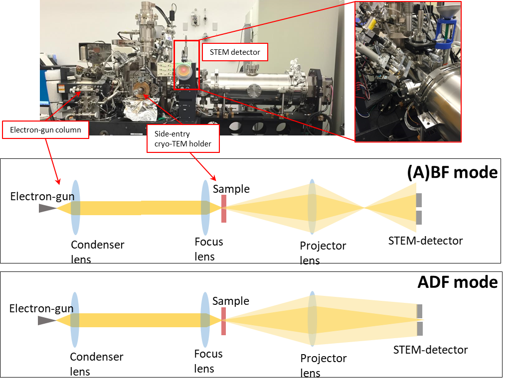
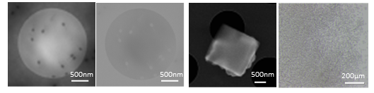
Figure 1. Top: Electron optics of the STEM modes in DMF4000 electron microscope developed. Bottom, left to right: ABF and ADF mages of bacteriophage T4, Lysozyme crystals images by electron and optical microscope
3.1.c Cryo-EM and MS studies of Shigella flexneri (Dr. Martin Cheung)
My primary research interests lie in the bacterial pathogen Shigella flexneri, the causative agent of the potentially fatal disease bacillary dysentery. The goal of my work is to elucidate the underlying mechanism by which S. flexneri rapidly invades target cells during infection. A key component of this process is the injectisome (also known as the type III secretion system), which enables the direct transfer of disease-causing proteins from S. flexneri into target cells. The tip complex (TC) is the outermost extracellular component of the injectisome and is thought to facilitate contact with target cells and to activate downstream regulatory processes once contact has been established. However, the precise means by which the TC achieves this is unknown.
To further our understanding of the TC, a multifaceted approach utilizing mass spectrometry and cryo-electron microscopy (cryo-EM) was employed. The data acquired by these techniques resulted in two key findings: (i) the TC comprises three distinct subunits with heterogeneous morphologies (Fig. 1a). Prior to this study, the TC had never been visualized to better than 20 nm resolution and was believed to be a homopentameric complex. By visualizing, for the first time, the TC to sub-nanometer resolution by cryo-EM, this work revealed the TC to be a trimeric complex; (ii) a hitherto unknown auxiliary complex binds to the TC upon activation of the injectisome during infection (Fig. 1b). A pore contiguous with the injectisome is known to be inserted into target cell membranes during infection. We believe this newly identified complex to be a pre-pore complex and may explain how the TC is able to interact with target cells. This work was funded in part by the AMED program and Grants-in-Aid for Scientific Research (KAKENHI). A paper based on these findings is currently being prepared.
(a) (b)


Figure 1. (a) Cryo-EM map of the TC. A trimeric complex comprising morphology distinct subunits can be seen. (b) Proposed pre-pore complex (green) that sits atop the TC (orange) following activation of the injectisome. Scale bar, 10 nm
3.1 d Electron holography studies of CdTe crystals and devices (Dr. Cathal Cassidy)
Following on from our publication on CdTe holography mean free path last year, we spent a lot of time to dig deeply into the microscope hardware parameters and underlying theory relating to mean free path in off-axis holography. We quantitatively measured the microscope optical parameters (such as biprism offset from image plane, and absolute electron deflection angles from specimen and biprism interactions). We developed a simple theoretical framework which explains the material factors which uniquely define the mean free path in electron holography. This work was heavily focused on electron holography, but serves as an important foundation for our planned studies of CdTe devices. A manuscript has been submitted and is presently under review. From December 2021, we established a CRA (Collaborative Research Agreement) with Acrorad – a company in Uruma specialized in development and manufacturing of CdTe devices. This will enable us to continue our research on holography of CdTe material & devices.
3.1 e Observing magnetic monopoles in spin ice (Dr. Ankur Dhar, Dr. Cathal Cassidy)
Based upon the thesis work of Dr. Ankur Dhar last year, in collaboration with Profs. Shannon & Jaubert, we prepared and submitted a manuscript on electron holography studies of magnetic monopoles. As Dr. Dhar has already graduated & departed for SLAC, Dr. Cassidy joined the project onsite at OIST. Our manuscript is available to view on arXiv, and publication activities are ongoing: https://arxiv.org/abs/2112.01362.
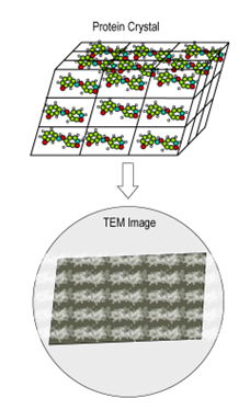
3.1 f Observation of Array of Molecule inside Protein Crystal (Dr. Tsumoru Shintake)
As well known, phase is missing in the recorded Bragg diffractions. In the history of X-ray protein crystallography, several important methods for phase determination were developed, and thus a huge number of 3D structure of proteins have been solved. However there are still many crystals unsolved, for example "twin crystals": protein crystal often creates twin crystals, which cannot be solved by X-ray crystallography, nor by Micro-ED.
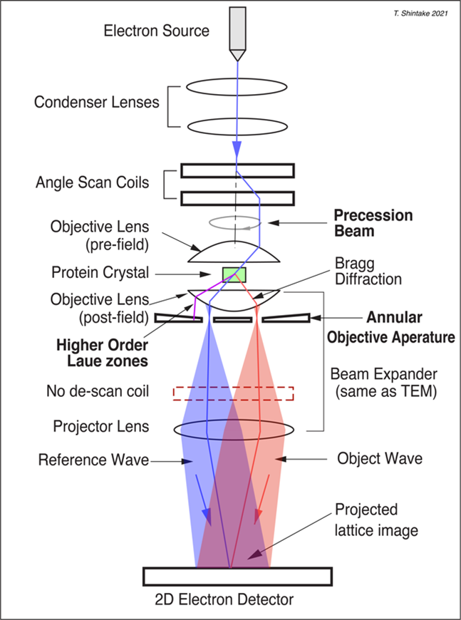
By utilizing the resolving power of today’s advanced TEM, if we should be able to observe inside the protein crystal and obtain image of the protein array. Thus, any type of crystal structure will be determined. However, this idea has been believed impossible, and many trials failed before. Additionally, there did not exist any theoretical basis which explains how to reconstruct a real image from Bragg diffractions through the lens.
Learning from the theory of the protein X-ray crystallography, Shintake discovered a new electron microscope principle, which will make it possible to directly observe array of molecule inside the protein crystals (https://arxiv.org/abs/2105.03850). The idea has been filed as provisional patent in 2021 (US Provisional Application 63/162, 866, March 2021).
After reviewing by Prof. Richard Henderson (Nobel prize in Chemistry 2017), we received positive recommendation. We started following R&Ds.
(1) Proof of principle experiment. JEOL Ltd. performed several experiments on their own TEM:JEM2100 at their laboratory. Operation of beam precession, filtering with annular slit. We created appropriate beam trajectory, while we have not yet tried crystal observation.
(2) Development of thin (100-300 nm) protein crystals, which is suitable for proposed microscopy. Dr. Yamashita suggested to crystal growing between two flat glasses.
We hope to obtain the first TEM image from protein crystal in 2022.
3.2 Ethanol Project Group
We started research on possible prevention method and cure for COVID-19 via ethanol inhalation, after the proposal by Dr. Shintake in 2020, " Possibility of Disinfection of SARS-CoV-2 (COVID-19) in Human Respiratory Tract by Controlled Ethanol Vapor Inhalation" (https://arxiv.org/abs/2003.12444). Under collaboration with Ishikawa Unit, we have been studying disinfection mechanism of influenza-A virus by exposure to ethanol (EtOH) in vitro, and treatment by ethanol vapor on the infected mice with influenza. We have very important results as follows:
(1) EtOH concentration required to disinfect the influenza virus is 20 %v/v at body temperature of 37 deg.C, which is much lower that the well-known value of 30%v/v at room temperature. We think the thermal energy contribute inactivation process together with EtOH chemical energy.
(2) By means of Cryo-TEM observations, we found conformational change of the surface proteins after inactivation with heat (>60 degree) or EtOH for 1 minute.
We continue to evaluate EtOH efficacy, and detail of the conformational change.
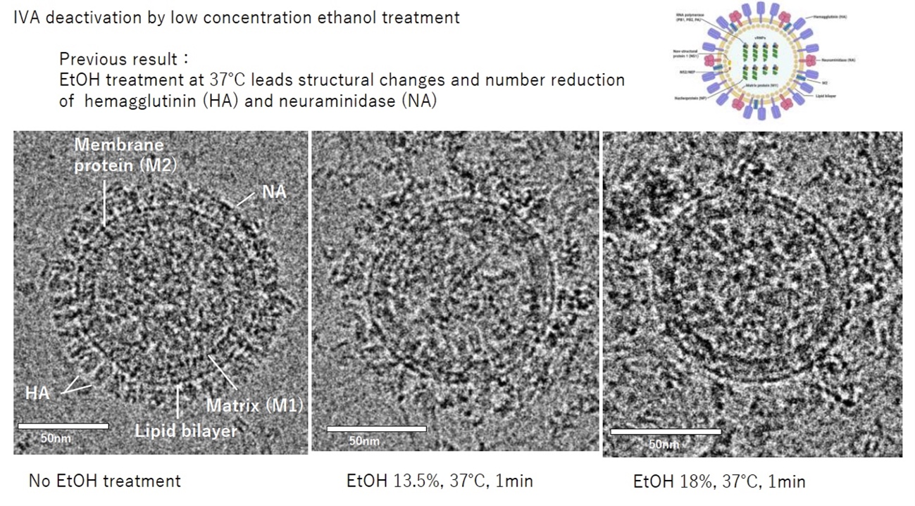
(Preliminary data) Conformational change on surface proteins on influenza-A virus after 1 minute exposure to EtOH.
3.3 Wave Energy Converter Group
OIST Wave Energy Project has been carrying out R&Ds to harness electricity from the moving water in the breaking wave zone since 2018, supported by Kokyo Tatemono Co. Ltd. in Tokyo, through their donation and contracted research.
However, what we learned from the project was that: the peak output-power driven by the breaking wave was fairly high, roughly 10 kW level for each 0.5 m diameter turbine/generator, while the average power, this is the most important parameter in renewable energy, was quite low, lower than 100 Watts unfortunately. We realized that there exists a fundamental problem: the breaking wave is chaotic phenomena, i.e., shape of the wave varies in time, also the breaking wave point moves back-and-forth. They are not predictable, thus probability of generating electricity was random and low after time averaged.
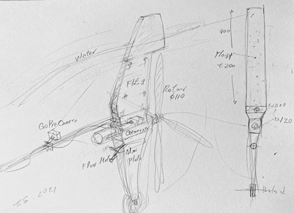
In order to overcome this problem, we decided to go deeper area, outside the wave breaking zone, where the moving water is smooth, like a sinuous motion of a pendulum oscillation, thus the output power becomes predictable. In the history of the wave energy development, no one had paid attention before in this area. OIST is the first place to utilize "Natural Oscillating Water Zone (OWZ)", which exists ubiquitously right near the wave breaking zone. New concept of wave energy device was proposed by Dr. Shintake in 2021 (September 30, 2021, US Provisional 2021-161724, Surface Ocean Wave Turbine)
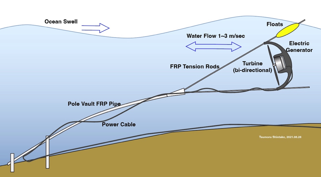
Figure 1. Submersible WEC located at OWZ: oscillating water zone
Through extensive studies via computer fluid simulations (CFDs), we confirmed existence of natural oscillating water zone near the breaking wave zone. We carefully analyzed wave permeance around the beach of Seragaki Port, where we have OIST Marine Center. We identified the OWZ, roughly 100 m away from the sea barrier, where the average water depth is 3 m. The computer simulation predicted maximum 2 m/sec flow speed, when the seasonal swells arrive from NW direction in Winter (November to March), from which we expected to harness 1 kW electricity by using 0.6 m diameter turbine.
We conducted CFD analysis on the beach of Kandooma Island Maldives, and confirmed OWZ.
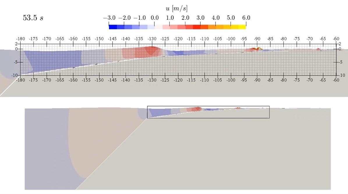
Figure 2. CFD simulation of the east side beach on Kandooma Maldives. Snapshot of the wave form, where the color indicates water flow speed. Wave height H = 1.2 m, T=10 sec.

Figure 3. Temporal water flow speed in time at x=-130 m, depth of 3 m, outside of the breaking wave zone. The forward/backward flows indicate OWZ phenomena
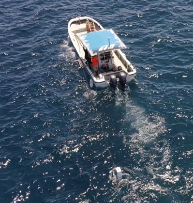
We have performed four times of ocean experiments on turbine, towing by boat at Maeda area.

07/29/2021 08/16/2021
Figure 4. The first demonstration model using half-scale generator with propellers from the powered paragliders. The turbine is located behind of the float, and thus this design is "down wind" type, which causes the shadow problem on the turbine, i.e., lowering the speed of water.
YAMAHA MOTOR CO.,LTD. jointed to our project, donated four HARMO (electric outboard motor) to OIST. We assembled one HARMO with three-blade turbine, and installed in seabed at 3 m deep, near drop-off in front of OIST Marine Science Station at Seragaki in April 2022.
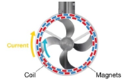
Figure 5. HARMO motor drive unit
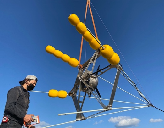
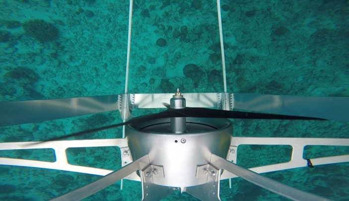
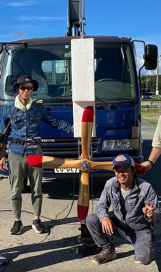
Figure 6. Submersible WEC using HARMO motor and three-blade turbine, which tested at Maeda port towing by motorboat.(02/17/2022)
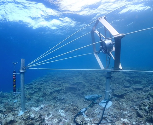
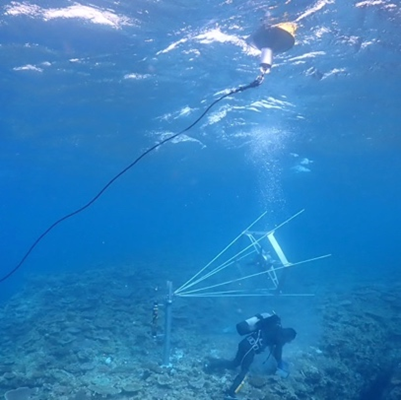
Figure 7. The first model of submersible WEC has been installed at the leaf-edge (3m deep) in front of the OIST Seragaki Marine Center. (04/09/2022). Hexagonal frame diameter is 1.2 m, and length of 2 m.
4. Publications
4.1 Journals
- T. Latychevskaia, C. Cassidy, T. Shintake, Bragg holography of nano-crystals” Ultramicroscopy230 113376; https://doi.org/10.1016/j.ultramic.2021.113376(2011)
- A. Dhar, L. Jaubert, C. Cassidy, T. Shintake, N. Shannon, Observing magnetic monopoles in spin ice using electron holographyhttps://doi.org/10.48550/arXiv.2112.01362
- C. Cassidy, M.T. Schreiber, M. Beleggia, J. Yamasaki, H. Adaniya, T. Shintake,Interpretation of mean free path values derived from off-axis electron holography amplitude measurements(under review)
- K.A. Pawitan, H. Takebe, H. Andrean, S. Misumi, J. Fujita and T. Shintake, Duct Attachment on Improving Breaking Wave Zone Energy Extractor Device Performance Energies 2021, 14(19), 6428; https://doi.org/10.3390/en14196428
4.2 Books and other one-time publications
Nothing to report
4.3 Oral and Poster Presentations
- M. Yamashita, J. Fujita, S. Taba and T. Shintake, Scanning electron microscopy on electrospray-generated biomolecular ions, The 77th Annual Meeting of the Japanese Society of Microscopy, 14-6 June (2021)
5. Intellectual Property Rights and Other Specific Achievements
- January 12, 2022, US 63/298,929, Proposal of Plane-Prallel Resonator Configuration for High-NA EUV Lithography
- October 12, 2021, Japan Filed Patent 2021-1667409,Nano-Flow Cytometry
- September 30, 2021, US Provisional 2021-161724,Surface Ocean Wave Turbine
6. Meetings and Events
Nothing to report
7. Other
7.1 Visitors
- 26 October: Delegation from Planning Department, Okinawa Prefectural Government
- 19 November: Delegation from Okinawa Promotion Bureau, Cabinet Office
- 25 November: Dr. Ingomar Lochschmidt and delegation from Advantage Austria, Austria Embassy
- 22 December: H.E. Midori Takeuchi, Ambassador of Japan to the Republic of Maldives
7.2 Media
- TBA