

FY2014 Annual Report
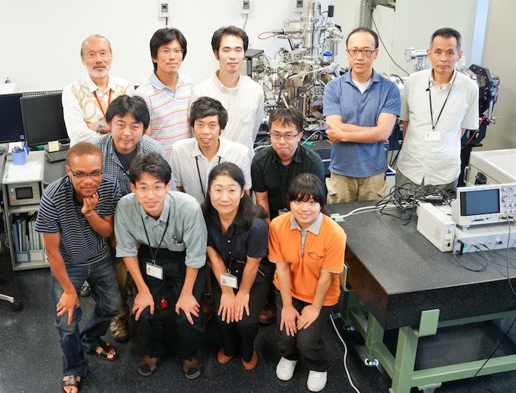
Quantum Wave Microscopy Unit
Professor Tsumoru Shintake
Abstract
1. R&D on diffraction electron microscope which uses the quantum wave interference associated with low energy electron as well as the technique of electron holography. With the use of a few tens of keV electron beam, and the phase-retrieval algorithms we are developing an electron microscope capable of imaging viral samples in sub-nanometer resolution without sample damage. The unit has so far developed an electron microscope, DMF4000, in collaboration with HITACHI High Tech Science, and the electron beam transfer system to image the diffraction pattern. In 2014 the unit has succeeded in producing the phase-retrievable holographic image.
2. We proposed a ocean-current turbine as a sustainable energy source in 2012. The turbine will convert the kinematic energy of an ocean-current into electricity. So far, in order to verify our ocean-current turbine design, our group conducted various towing tests and mooring experiments by scale models. Now, we are working on developing a 10 kW model to perform a feasibility study by tidal current. In FY 2014, we conducted two R&D items. First, we installed an observation floating body to measure a tidal current and the floating body motion. Secondly, we evaluated the turbines that were designed by us. The performance was measured at a circulating water channel.
Staff
- Dr. Katsutoshi Shirasawa, Staff Scientist
- Dr. Hidehito Adaniya, Postdoctoral Scholar
- Dr. Teruhisa Hirai, Staff Scientist
- Dr. Masao Yamashita, Postdoctoral Scholar
- Dr. Martin Philip Cheung, Postdoctoral Scholar
- Hideki Takebe, Technician
- Jun Fujita, Technician
- Junichiro Minami, Technician
- Chola Kalale, OIST PhD Student
- Ayumi Shimojima, Research Administrator
Collaborations
- Theme: Development of the ocean current turbine
- Type of collaboration: Joint research
- Researchers:
- Professor, Dr. Hidetsugu Iwashita, Hiroshima University
- Student, Kohei Tokunaga, Hiroshima University
-
Theme: Development of novel specimen grids for Transmission Eloctron Microscopy (TEM)
- Type of collaboration: Joint research
- Researchers:
- Professor, Dr. Jun Mizuno, Waseda University
- Student, Hiroyuki Kuwae, Xu Bingyang, Waseda University
Activities and Findings
Electron microscope group
Conceptual design of Diffraction electron microscope
In principle, by applying the method of iterative phase retrieval on coherent diffraction data, one can determine the phase of the diffraction beam, and reconstruct the real image of the sample object (Fig.1). This process works correctly if the object has a two dimensional structure. The phase of the diffraction beam from an object of three dimensional structure, however, cannot be determined uniquely. We have found that we may solve this problem by introducing holographic image reconstruction. To perform this experiment on biological sample, one may use an electron beam in low energy, in the range of a few keV to a few tens of keV, to prevent the beam damage on the target sample. And the sample needs to be a thin enough to allow for the transmission of the diffracting electrons.
Development of the electron diffraction microscope
Prior to 2014 we have developed an electron microscope with focused ion beam, DMF4000, which is capable of SEM imaging and FIB etching. We also have developed the electron beam transport system which is to obtain the diffraction patterns of scattered electron of low energy electron beam, smaller than a few keV energy. The essential features of this system are the 2dimensional detector of 8k x 8k COMOS sensors and electron optics system, which make possible to measure the diffraction pattern produced by large Bragg angles of atomic scale spacing. The unit has also been developing a technique of FIB-etching the test target smaller than a few hundred nanometers, compatible to the actual viral size, on metal coated TEM grids.
With the test target the unit has developed the techniques of producing the holography image in a few nanometers resolution. It was found that in addition to the properties of electron beam, such as coherency and stability, the physical properties of the surface of the TEM grid play a major role for the re-constructability of holography and high resolution. The development of the reconstruction technique of the real image from the holography pattern in a stable manner is actively under progress.
To facilitate the holographic diffraction imaging on insulating biological specimens, we have been optimizing the specimen design with the following technical considerations in mind; 1) specimen transparency to low energy electrons, 2) specimen charge-up upon electron beam irradiation, and 3) changes in specimen morphology by vacuum dehydration. To address 1) and 2), we have developed the graphene support film for biological specimens, which is only one carbon atom thick and is highly conductive. We have established a fabrication scheme for the graphene TEM grids so far. Regarding 3), we have developed on virus and DNA specimens a freeze drying technique, which preserves the fine sample structures to a few nm resolution. Future work will address the implementation of the common technique of ice embedding for the low energy electron imaging on freeze-hydrated specimens.
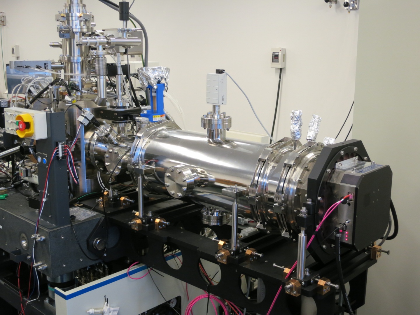
Figure 1.: OIST electron microscope; from left to right, DMF4000, electron diffraction spectrometer, F816 CMOS detector.
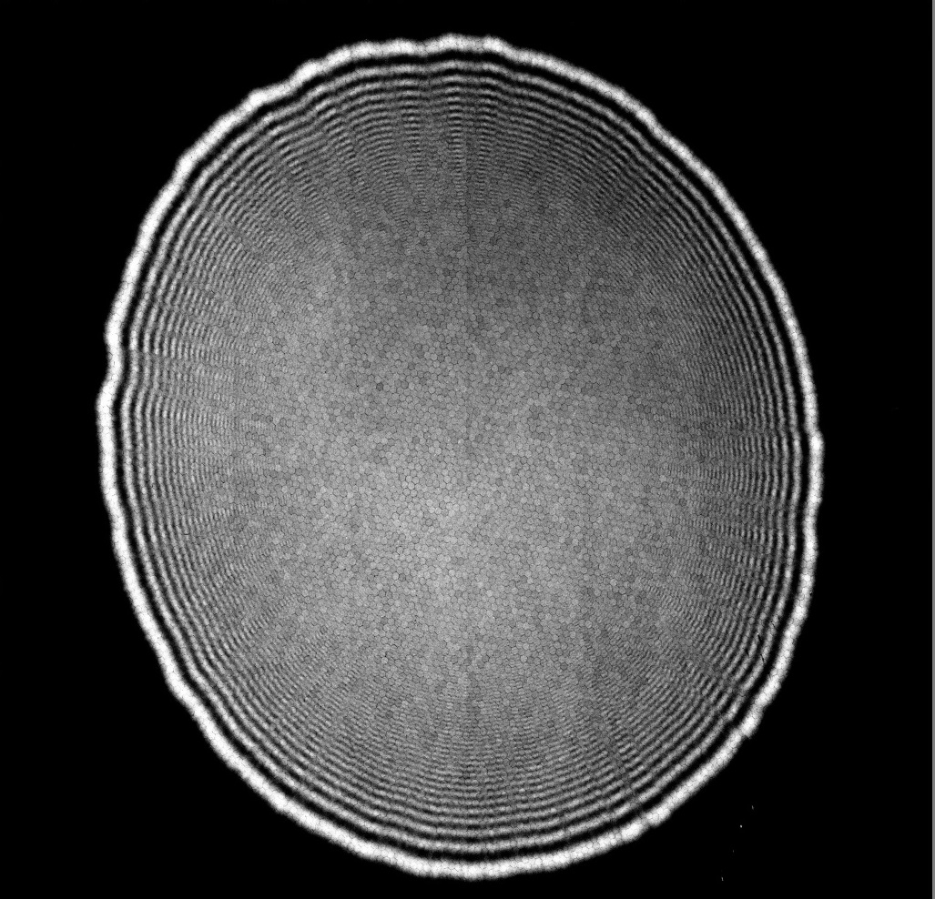
Figure 2.: Fresnel fringe pattern of 30 micrometer aperture by 10keV electron beam, showing high coherency of the electron beam. The image is obtained by F816 CMOS detector.
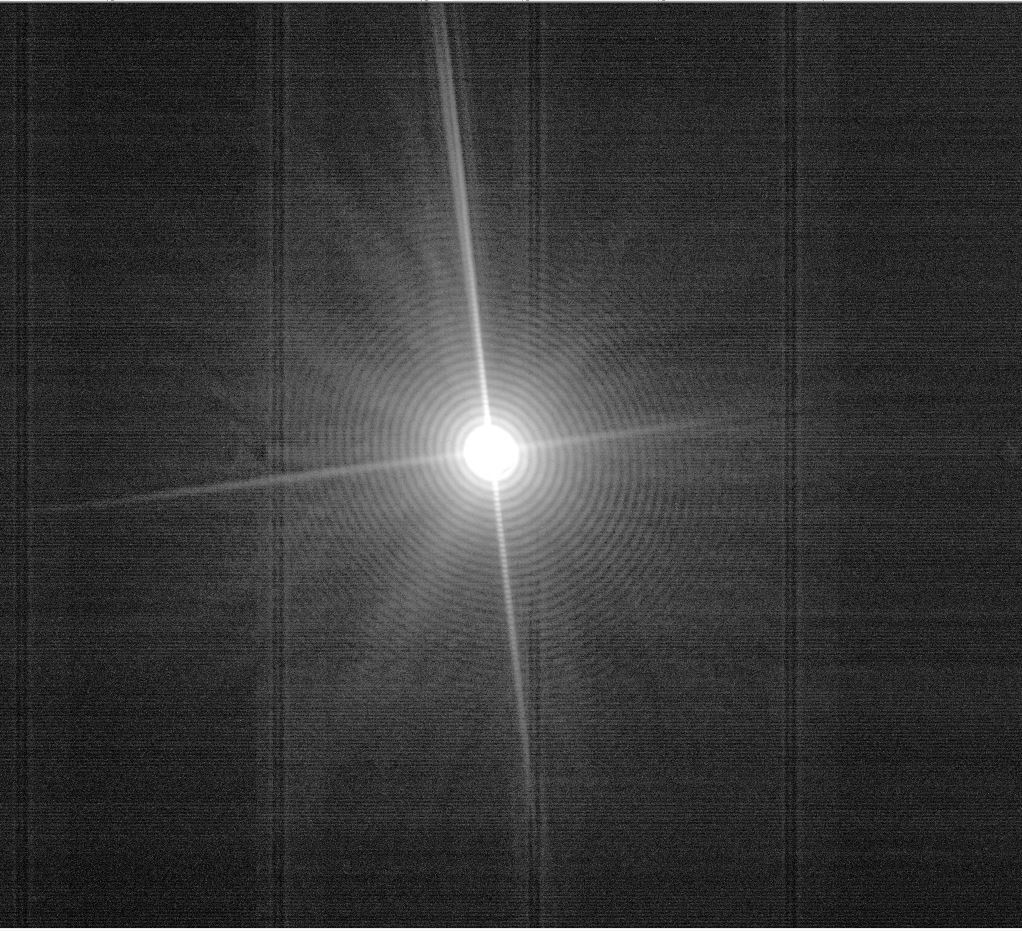
Figure 3.: Diffraction image from our test target taken F816 CMOS detector. The resolution in the holography image from this diffraction is estimated to be 3~4 nanometers.
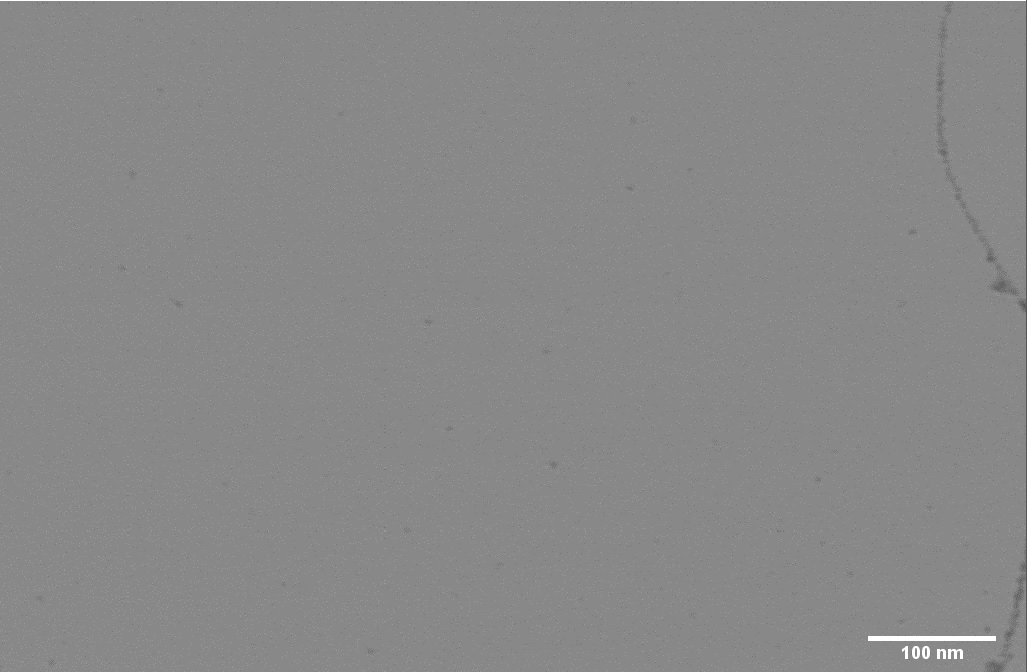
Figure 4.: 10 kV scanning transmission electron microscope (STEM) image of the graphene support film. The two lines in the image correspond to the boundary of two graphene domains but otherwise the graphene shows no texture.
Ocean Power Generation Group
Observation floating body
Our ocean-current turbine will work in a middle layer of an ocean current to avoid ocean waves. In FY 2014,in order to obtain real tidal current data, we installed the observation floating body into the northern sea of Okinawa. The schematic diagram is shown in Fig. 1.Th e floating body is equipped with depth meters, thermometers and accelerometers. On the sea surface, the communication buoy is working to send the measured data to our database. In addition, the buoy has an ADCP (Acoustic Doppler Current Profiler) to measure current speed, direction and profile. We will analyze the measured data to optimize our turbine performance and mooring system.
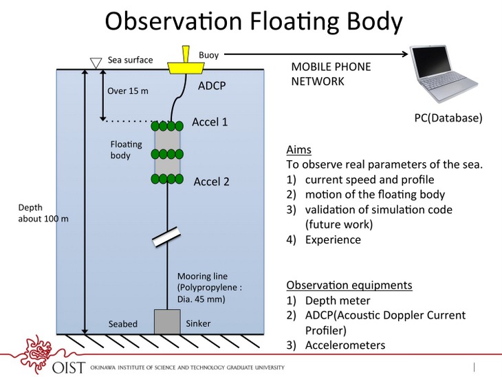
Figure 1: System diagram of the observation floating body.
Performance test of scale model turbines
We designed three scale model turbines to investigate the power performance. The scale models were processed by a 5-axis CNC machine. The material is aluminum and the diameter is 300-mm. The photograph of the turbine is shown in Fig. 2. The performance was evaluated at a circulating water channel of the West Japan Fluid Engineering Laboratory Co., LTD. We measured the torque, rotation speed and thrust force. The results are in good agreement with the design values. The maximum power coefficient (Cp) was 0.42. As the next step, we are going to design more efficient turbine and optimize the turbine structure for ocean currents.
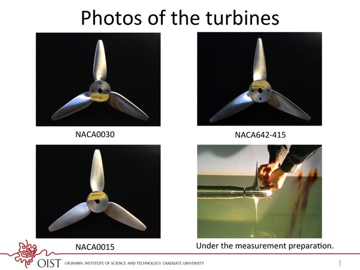
Figure 2: Photograph of the scale model turbines.
4. Publications
4.1 Journals
- Shirasawa, K., Shintake, T., Development of an Ocean Current Turbine at OIST, in Bulletin of the Society of Sea Water Science, Japan, Volume 68, Number 5, PP282-206 (2014).
- Shintake, T., Building a Sustainable World, in Scientific American Special issue published in partnership with ALSTOM, Supplement of the french edition no447, p41 (2015).
4.2 Books and Other One-Time Publications
Nothing to report
4.3 Oral and Poster Presentations
- Shintake, T., SACLA X-RAY FEL BASED ON C-BAND TECHNOLOGY (GERSCH BUDKER PRIZE LECTURE), in 5th International Particle Accelerator Conference (IPAC14), International Congress Center Dresden, Germany (2014).
- Shintake, T., SACLA Hard X-ray Free Electron Laser Based on Normal Conducting Accelerator Technology, in 12th International School and Symposium on Synchrotron Radiation in Natural Science (ISSRNS14), Conference center at Hotel BOSS, Warsaw, Poland (2014).
- Shintake, T., Proposal of Rotating Blade Wave Energy Converter Installed in the On-Shore Wave Breaking Zone (Possibility of Intelligent Tetrapod), in Ocean Energy Symposium 2014, Institute of Ocean Energy, Saga University (2014).
- Shirasawa, K., Shintake, T., Technological development of ocean current power generation in OIST, in Seawater Society of Japan the 65th Annual Symposium, Tiruru, Okinawa Center for Gender Equality, Okinawa, Japan (2014).
- Shirasawa, K., Shintake, T., Minami, J., Tokunga K., Iwashita, H., Hashizume, Y., The mooring test by the miniature model of OIST ocean current generator, in The Japan Society of Naval Architects and Ocean Engineers (JASNAOE) 2014 Annual Spring Meeting, Sendai International Center, Miyagi, Japan (2014).
- Shirasawa, K., Shintake, T., Tokunga K., Iwashita, H., Development of an ocean current turbine at OIST, in International symposium on ocean renewable energy technologies, Kyushu University, Kasuga, Fukuoka, Japan (2014).
- Shirasawa, K., Tokunga K., Iwashita, H., Shintake, T., Submerged Hydraulic Turbine for Deep Marine Current as a Electric Power Generator, in Grand Renewable Energy 2014 International Conference and Exhibition, Tokyo Big Sight, Tokyo, Japan (2014).
- Hirai, T.,Structural analysis of human erythrocyte AE1 (band 3),in The 76th Annual Meeting of the Japanese Society of Hematology, Osaka International Convention Center, Osaka (2014).
5. Intellectual Property Rights and Other Specific Achievements
Nothing to report
6. Meetings and Events
6.1 Seminars
Title: Coherent Diffractive Imaging of Cells and Macromolecular Complexes at SPring-8 and SACLA
- Date: April 9, 2014
- Venue: OIST Campus Lab1 Meeting Room C016
- Speaker: Mr. Marcus Gallagher-Jones, Ph.D. student, Liverpool University
Title: Multi-Purpose CMOS Cameras for Electron and X-Ray detection; Sensitivity, Resolution, Dynamic Range, Speed
- Date: May 15, 2014
- Venue: OIST Campus Lab1 Meeting Room C015
- Speaker: Mr. Hans Richard Tietz, Managing Director, Tietz Video and Image Processing Systems GmbH, Germany
Title: Future High-Energy Colliders / Charting the Unknown
- Date: February 18, 2015
- Venue: OIST Campus Lab1 Meeting Room C015
- Speaker: Dr. Frank Zimmermann, Senior Scientist at CERN (Switzerland)
Title: Project of a 3 GeV High Brilliant SYnchrotron Light Source, "SLiT-J"
- Date: March 30, 2015
- Venue: OIST Campus Lab1 Meeting Room C015
- Speaker: Dr. Hiroyuki Hama, SLiT-J Office, Electron Photon Research Center, Tohoku University
7. Other
Nothing to report.