

Seminar on "Structural and functional studies of the Type III Secretion system of Shigella flexneri" by Dr. Martin Cheung
Date
Location
Description
Quantum Wave Microscopy Unit would like to invite you to a seminar by Dr. Martin Cheung.
Time/Date: 13:30 - 14:30 Wednesday, October 30
Venue: Lab 1 Meeting Room C015 (Level C)
Speaker: Martin Cheung, Post-doctoral research assistant, University of Bristol
Seminar Abstract:
The Type III Secretion System (T3SS) plays a pivotal role in the virulence of many Gram-negative bacteria. Numerous genera of bacteria rely on this system for virulence and strains with dysfunctional systems have attenuated virulence. Bacteria utilising the T3SS include those involved in human, animal and plant pathogenesis. Shigella flexneri is the bacterial cause of shigellosis, the most acute form of bacterial dysentery. A global endemic, shigellosis is of particular burden in the developing world where it accounts for 75% of all diarrhoea related deaths.
The widespread use of the T3SS by pathologic bacteria reflects its importance in virulence and it is therefore not difficult to see the T3SS as a potential target for new antimicrobials and vaccines. A “molecular syringe”, the T3SS is a 3.5MDa transmembrane complex that allows for the direct transfer of proteins from the bacterial cytoplasm into the host cell. The three main components of the T3SS are (i) the base that spans both bacterial membranes, (ii) needle, which emanates 50nm from the cell surface and (iii) tip complex (TC) that forms a pore in the host cell membrane. To aid the rational design and discovery of inhibitors of T3SSs, much research is being invested in defining the structure of the T3SS and relating this structural knowledge to its function. Of particular interest is the activation mechanism and signal transduction through the system.
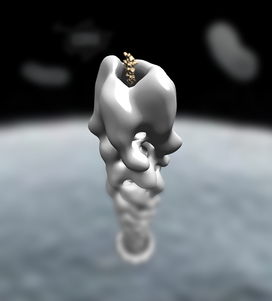
During my PhD, I used electron microscopy to study the three key components of the T3SS. Utilitsing negative stain EM, we were able to produce 20 - 25 Å density maps of the TC in wild-type and activated states, revealing for the first time its asymmetrical architecture. We were also able to produce a 3D reconstruction of the needle to 7 Å, allowing for direct visualisation of how the needle subunit is able to polymerise into an extended structure. The new structural information gained during my PhD has allowed us to develop theories into how the T3SS functions and has paved the way for the development of drugs specifically targeting this system.
Subscribe to the OIST Calendar: Right-click to download, then open in your calendar application.