FY2016 Annual Report
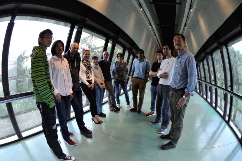
Optical Neuroimaging Unit
Associate Professor Bernd Kuhn

Photo by Emiko Asato
Abstract
In the FY 2016 the Optical Neuroimaging Unit continued several imaging projects. Most research projects focused on imaging neuronal activity in the mouse brain.
1. Staff
- Dr. Bernd Kuhn, Associate Professor
- Dr. Akihiro Funamizu, Postdoc (shared with Doya Unit)
- Dr. Christopher J. Roome, Postdoc
- Dr. Sigita Augustinaite, Postdoc
- Dr. Shinobu Nomura, Postdoc
- Ray X. Lee, Ph.D. student
- Neil Dalphin, Ph.D. student
- Leonidas Georgiou, Ph.D. student
- Dr. Kazuo Mori, Technical Assistant
- Hiroko Chinone, Research Unit Administrator
2. Collaborations
2.1 Neural substrate of dynamic Bayesian inference in the cerebral cortex
- Dynamic Bayesian inference allows a system to infer the environmental state under conditions of limited sensory observation. We use in vivo two-photon calcium imaging and probabilistic population decoding to show that cortical neurons in layers 2, 3, and 5 implement the fundamental features (prediction and updating) of Dynamic Bayesian inference.
- Type of collaboration: Joint research
- Researchers:
- Professor Dr. Kenji Doya, Neural Computation Unit, OIST
- Dr. Akihiro Funamizu, Neural Computation Unit and Optical Neuroimaging Unit, OIST
3. Activities and Findings
3.1 Neural substrate of dynamic Bayesian inference in the cerebral cortex
Dynamic Bayesian inference allows a system to infer the environmental state under conditions of limited sensory observation. Using a goal-reaching task, we found that posterior parietal cortex (PPC) and adjacent posteromedial cortex (PM) implemented the two fundamental features of dynamic Bayesian inference: prediction of hidden states using an internal state transition model and updating the prediction with new sensory evidence. We optically imaged the activity of neurons in mouse PPC and PM layers 2, 3 and 5 in an acoustic virtual-reality system. As mice approached a reward site, anticipatory licking increased even when sound cues were intermittently presented; this was disturbed by PPC silencing. Probabilistic population decoding revealed that neurons in PPC and PM represented goal distances during sound omission (prediction), particularly in PPC layers 3 and 5, and prediction improved with the observation of cue sounds (updating). Our results illustrate how cerebral cortex realizes mental simulation using an action-dependent dynamic model. This paper was published in 2016.
3.2 Voltage imaging with two-photon microscopy
Many neurons have elaborate dendrites with a wide variety of ion channels governing their electric properties. Unfortunately, it is very difficult to record the voltage in dendrites due to their fine structure. At the same time dendritic voltage is a key to understand neuronal information processing and therefore also to understand brain function.
We develop tools to measure dendritic voltage changes in vivo. We use the pure electrochromic voltage sensitive dyes ANNINE-6 (Kuhn & Fromherz 2003) and ANNINE-6plus (Fromherz et al 2008). Pure electrochromic means that the change of fluorescence only depends on the electric field over the dye molecule and that there are no other, slower effects modulating the spectra.
We label single neurons in vivo through an access port in the chronic cranial window (Roome & Kuhn 2014). By excitation at the red edge of the a absorption spectrum we can reduce phototoxicity and increase voltage sensitivity dramatically (Kuhn et al 2004).
After filling a cell we can simultaneously electrically record from the soma and image dendritic voltage and calcium changes in awake animals. We study Purkinje neurons in the cerebellum. We can observe supra and subthreshold voltage changes.

3.3 Imaging thalamo-cortical interaction
Sensory information is relayed through the thalamus to cortex. At the same time, cortex is regulating thalamic activity by driving and modulatory feedback. We image layer 6 pyramidal neurons in primary visual cortex which send a feedback signal to primary visual thalamus during different behavioral states. This will allow us to better understand the thalo-cortical loop.
3.4 Imaging PKA activity in vivo
Protein kinase A (PKA) is an important enzym in the signaling cascade connecting neuronal activity and expression and modulation of proteins. We use genetically encoded indicators to image PKA activity in vivo.
3.5 Post-traumatic behavioral development in adult mice
PTSD is one of the most prevalent mental health disorders and has received serious clinical and research attention (DSM-III, 1980;DSM-5, 2013). Still, it remains as one of the most poorly understood and controversial presentations of psychiatric disorders. Here, we explore how bonding explains post-traumatic behavioral development in adult mice.
3.6 pH and Ca2+ measurement in nanopores of protein crystals
Protein crystals can be considered nano porous materials. We measure the conditions inside the nano pores with the help of molecular probes. We use pH-sensitive dyes, load them into the crystals, and record pH changes in response to pH changes of the bath. We find differences between the pH of the bath and the nano pores. We try to find a model that explains this difference.
4. Publications
4.1 Journals
- A. Funamizu, B. Kuhn, and K. Doya* (2016) Neural substrate of dynamic Bayesian inference in the cerebral cortex, Nature Neuroscience 19: 1682-1689
4.2 Books and other one-time publications
Nothing to report
4.3 Oral and Poster Presentations
-
C.J. Roome, B. Kuhn (2017) In’s and Out’s of Neuronal Computation: Simultaneous Dendritic Voltage and Calcium Imaging and Somatic Recording from Purkinje Neurons in Awake miceGordon Conference ‚Dendrites‘, Lucca, Italy
-
R.X. Lee, G.J. Stephens, B. Kuhn (2017) Dynamics of somatic and dendritic populations in cerebral cortex during spontaneous movement onset, Computational and Systems Science (Cosyne) 2017, Salt Lake City, USA
-
K. Mori, B. Kuhn (2016) Optical measurement of diffusion and pH in nanopores of protein crystals, Annual Meeting of the Biophysical Society of Japan, Tsukuba, Japan
-
C.J. Roome, B. Kuhn (2016) In’s and Out’s of Neuronal Computation: Simultaneous Dendritic Voltage and Calcium Imaging and Somatic Recording from Purkinje Neurons in Awake mice, Society for Neuroscience Meeting, San Diego, USA
-
C.J. Roome, B. Kuhn (2016) In’s and Out’s of Neuronal Computation: Simultaneous Dendritic Voltage and Calcium Imaging and Somatic Recording from Purkinje Neurons in Awake mice, Champalimaud Neuroscience Symposium, Champalimaud Foundation, Lisbon, Portugal
-
A. Funamizu, B. Kuhn, K. Doya (2016) Neural substrate of dynamic Bayesian inference in posterior parietal cortex, The 39th Annual Meeting of the Japan Neuroscience Society, Yokohama, Japan
-
C.J. Roome, B. Kuhn (2016) Exploring input-output relations of neurons in vivo, The 39th Annual Meeting of the Japan Neuroscience Society, Yokohama, Japan
-
R.X. Lee, G.J. Stephens, B. Kuhn (2016) Neuronal Interaction Dynamics in the Neocortex during Spontaneous Locomotion Onsets, The Origin of Consciousness, ELSI, Tokyo Institute of Technology, Tokyo, Japan
-
The Seventh International Neural Microcircuit Conference: Recent advances in the analysis of cortical microcircuits “Neural substrate of dynamic Bayesian inference in the cerebral cortex”, Organizer: Prof. K. Kobayashi, Prof. Y. Kubota, (December 8-10, 2016), Okazaki Conference Center, Okazaki, Japan
-
Photonics Meeting, “Imaging neuronal activity in awake mice”, Organizer: Prof. K. Wakita, Chiba Institute of Technology (December 2-3, 2016), Okinawa Seinenkaikan, Naka, Japan
-
Biophysical Society Meeting of Japan Symposium “Advances in imaging neuronal activity: New tools and applications”, Exploring input-output relations of neurons in awake mice (November 25, 2016), Tsukuba, Japan
-
OIST-JST Presto Joint Symposium on Frontiers in Optics and Photonics (October 31, 2016), Organizer: Frontier-PJ, JST presto, OIST, OIST, Japan
-
OIST Mini Symposium “Science and Technology at the Interface of Bio-Nano-Systems: Challenges and Opportunities” (April 25-27, 2016), Organizer: Prof. Amy Shen, OIST, Japan
5. Intellectual Property Rights and Other Specific Achievements
Nothing to report
6. Meetings and Events
6.1 Workshop: Okinawa Computational Neuroscience Course 2016
- Date: June 13- June 30, 2016
- Venue: OIST Seaside House
- Organizers: Drs. E. De Schutter, K. Doya, J. Wickens
- Speaker (among others): Bernd Kuhn
- Title 1: Voltage-gated channels and the Hodgkin-Huxley model of neuronal activity
- Title 2: Functional Optical Imaging Methods
6.2 Onna-son/OIST Children’s School of Science 2016
- Date: August 22-26, 2016
- Venue: Fureai Taiken Center
- Speaker (among others): Bernd Kuhn
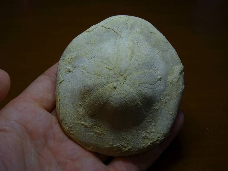
- Topic: Palaeontology of Okinawa for Grade 1-3

7. Other
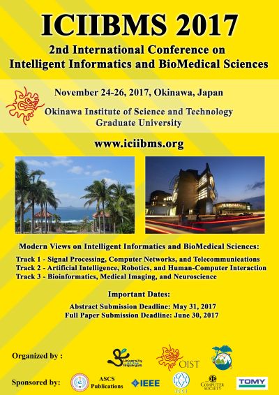
7.1 Planning of ICIIBMS 2017
- The Second International Conference on Intelligent Informatics and BioMedical Sciences (ICIIBMS) is in the planning phase.
- Date: November 24-26, 2017
- Venue: OIST
- Organized by researchers of the University of the Ryukyus, the Okinawa National College of Technology, and OIST


