FY2022 Annual Report
Quantum Wave Microscopy Unit
Professor Tsumoru Shintake
Abstract
Electron microscopy group
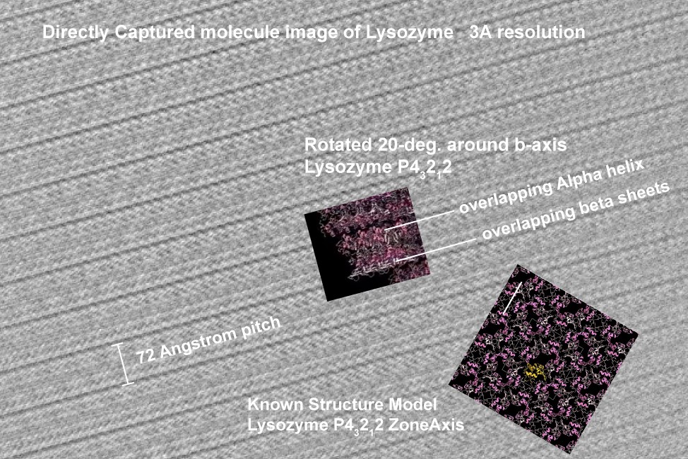
We successfully captured a high-resolution image of molecule array inside protein crystal by our new electron microscopy based on OIST invention(https://arxiv.org/abs/2105.03850). It was believed impossible to see inside the protein crystal at atomic resolution before.
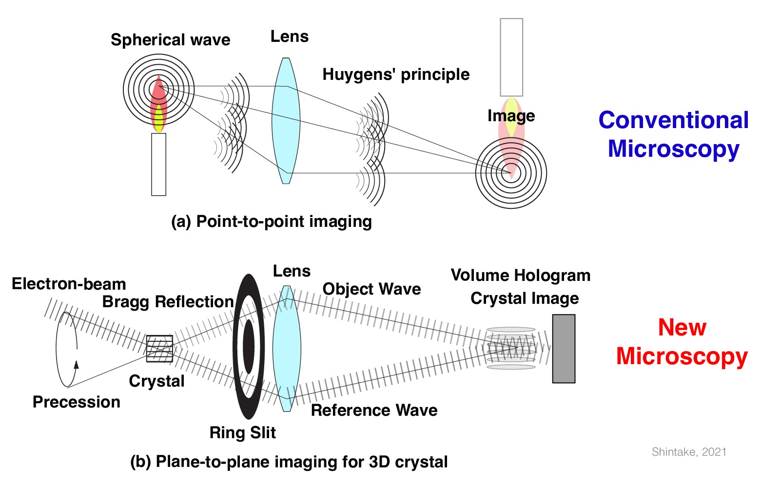
In the conventional optics (upper figure), the light wave emitted from a point source in the candle propagates through lens and focus into a point at downstream. This is point-to-point imaging system. Optical and electron microscopes are same in this concept. On the other hand, our new microscope (lower figure) works as plane-to-plane imaging. The reflected wave from the crystal goes through the ring slit and focus at downstream and overlap with illumination beam, create interference fringes. Quantum mechanical probability function tells us the electron wave takes maximum on the lattice plane inside the crystal, which is imaged on the detector. What we get on the detector is 2D projection image of the crystal. Because of the ring slit, the spherical aberration of the focusing lens inside the electron microscope is effectively eliminated, and thus it may reach atomic resolution. In 2022, we upgraded the existing electron microscope (JEOL CRYO ARM300) at Prof. Keiichi Namba laboratory at Osaka University, where we placed the ring slit inside lens. Dr. M. Yamashita and Dr. H. Adaniya studied sample preparation method suitable for this microscopy at OIST. The first experiment was performed at the end of March 2023, and we took the image shown above. We were surprised about beautiful array of molecules, and it was already 3 Angstrom resolution. In the next step, we will develop software to assemble multiple images from different orientation and create 3D protein structure. There will be many applications, in biology, in material science, or molecule-based technology development.
We published our work on the interpretation of mean free paths in electron holography in the journal Micron this year. We renewed our Collaborative Research Agreement (CRA) with local company Acrorad, and additionally extended the agreement to include Siemens Healthineers as one of the collaboration partners. We continued our experimental efforts on electron holography of magnetic monopoles, based on the thesis work of Dr. Ankur Dhar, and hope to re-submit the associated manuscript soon.
New telecentric optics for EUV lithography was proposed by Shintake, in January 2022 (https://arxiv.org/abs/2201.06749), which will solve the fundamental problem (mask 3D effect) in the current EUV photolithography. If it works, 5 nm or even narrower line process will be realized in the semiconductor device fabrication in future. It has been filed as US provisional patent.
Ethanol project group
The COVID-19 pandemic affected all of our life. We need to establish effective clinical care for the future. It should be based on a fundamental understanding of molecule biology. We study how the safety chemical of ethanol (EtOH) alters the entry protein by observing under cryo-electron microscopy at the atomic level, how the virus titler may be reduced by EtOH on model cells in vitro, and how much improvement can we see in infected mice. We have been studying the influenza virus because we can handle it in P2-level bio-safety control. SARS-COV2 (COVID-19) and the influenza virus are closely related, i.e., they are both envelop viruses, that cause the same respiratory illness and pandemic.
Summary
(1) We are investigating ethanol vapor inhalation treatment as a new clinical care.
(2) We found ethanol vapor using 50% v/v concentration reduced influenza viruses in the lung and could improve survival in the mouse model, which has been published in The Journal of Infectious Diseases.
(3) We are planning clinical trials to see how EtOH vapor may apply to humans. It will be conducted in hospitals around Okinawa.
This study was initially started by our theoretical study in 2020 (arXiv:2003.12444), and performed in collaboration with Ishikawa Unit at OIST.
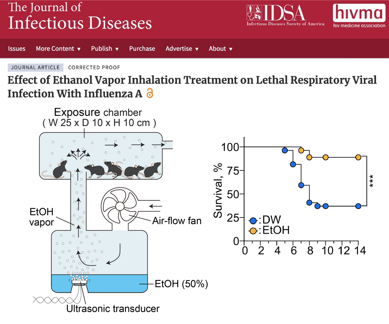
Figure of Development on Ethanol Inhalation Treatment. Joint research with the Shintake Unit and Ishikawa Unit has shown that mice treated with ethanol vapor are protected from lethal respiratory infections caused by the influenza A virus. The device shown on the left was developed and ethanol treatment was performed, resulting in improved survival rates as shown on the right. This treatment may also be effective against similar viral infections such as the new coronavirus. The results were published in The Journal of Infectious Diseases.
Wave energy project group
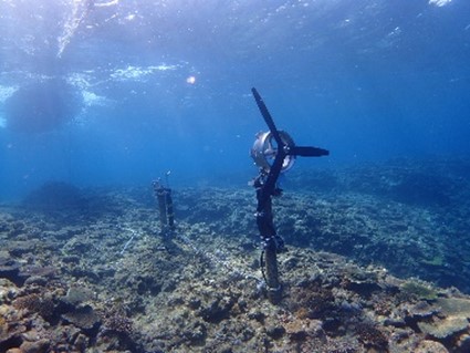
New concept of wave energy device was proposed by Shintake in 2021 (September 30, 2021, US Provisional 2021-161724, Surface Ocean Wave Turbine) and YAMAHA MOTOR CO.,LTD. jointed our project, donated four HARMO (electric outboard motor) to OIST. We assembled one HARMO with three-blade turbine, and installed in seabed at 3 m deep, near drop-off in front of OIST Marine Science Station at Seragaki in April 2022.
1. Staff
- Dr. Cathal Cassidy, Staff Scientist
- Dr. Masao Yamashita, Staff Scientist
- Dr. Ryo Kanno, Staff Scientist
- Dr. Takeshi Mise, Staff Scientist
- Dr. Hidetoshi Adaniya, Research Unit Technician
- Seita Taba, Research Unit Technician
- Jun Fujita, Research Unit Technician
- Shuji Misumi, Research Unit Technician
- Maximilian Vogl, Research Unit Technician
- Hideki Takebe, Specialist
2. Collaborations
2.1 Imaging magnetic monopoles in spin ice
- Type of Collaboration: Joint Research
- Researchers :
- Dr. Ankur Dhar, SLAC
- Dr. Ludovic Jaubert, CNRS Bordeaux
- Prof. Nic Shannon, OIST
- Dr. Cathal Cassidy, OIST
2.2 Application of electron holography and related analysis techniques to investigate electrostatic potential distributions at metal-CdTe interfaces
- Type of Collaboration: Collaborative Research Agreement (CRA)
- Researchers :
- Dr. Cathal Cassidy, OIST
- Tamami Unten, Acrorad
- Dr. Miguel Labayen de Inza, Siemens Healthineers
2.3 Observation of Array of Molecule inside Protein Crystal,Based on OIST Invention
- Type of Collaboration : Joint Research
- Researchers :
- Prof. Tsumoru Shintake, OIST
- Dr. Masao Yamashita
- Dr. Hidehito Adaniya
- Dr. Ryo Kanno
2.4 Protein Crystal Imaging with Precession TEM
- Type of collaboration : Joint Research
- Researchers :
- Prof. Tsumoru Shintake, OIST
- Dr. Masao Yamashita
- Dr. Ryo Kanno
- Seita Taba
- Prof. Keiichi Namba, Osaka University
- Dr. Fumiaki Makino, Osaka Universitry
2.5 Research on Ethanol Treatment on Infected Influenza A Mice
- Type of collaboration: Joint research with Ishikawa Unit
- Researchers :
- Prof. Tsumoru Shintake
- Prof. Hiroki Ishikawa
- Dr. Miho Tamai
- Dr. Melissa Matthews
- Dr. Masao Yamashita
- Dr. Ryo Kanno
- Dr. Takeshi Mise
- Seita Taba
- Jun Fujita
2.6 OIST Wave Energy Converter Project (WEC)
- Type of collaboration: Contracted Research
- Researchers :
- Prof. Tsumoru Shintake, OIST
- Shuji Misumi
- Jun Fujita
- Hideki Takebe
- Koma Ariga, YAMAHA MOTOR CO.,LTD.
- Atsushi Murashima, YAMAHA MOTOR CO.,LTD.
- Shintaro Futagawa, YAMAHA MOTOR CO.,LTD.
- Hikaru Kamimura, Kokyo Tatemono Co.,Ltd.
- Hiroshi Shimano, Kokyo Tatemono Co.,Ltd.
- Hamish Taggart, OPM Ocean Paradise Maldives
3. Activities and Findings
3.1 Electron microscopy group
3.1.a Development of electrospray sample deposition system(Dr. Masao Yamashita)
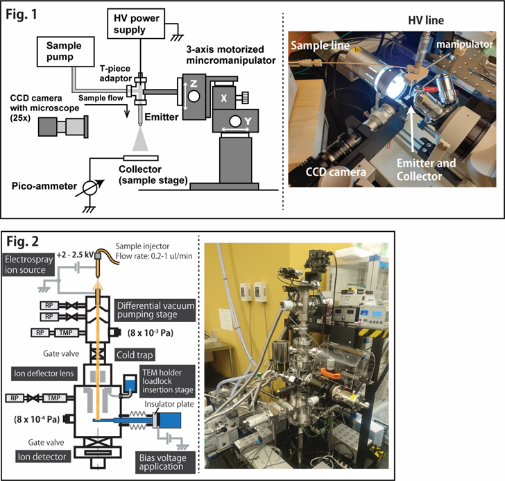
Our unit has been working on developing a new biospecimen preparation system for EM based on electrospraying (ES) technology. We have long been focusing on such application of ES since it can potentially be used to generate desolvated “naked” biomolecules, which is logically the most ideal form for specimens to be imaged by TEM/STEM at low voltages. However, this kind of application of ES is not common as it is only applicable to those samples that can meet very specific and stand with rather harsh conditions. For this reason, ES has rarely been applied to large biological macromolecules and, as a result, how well biomolecular structures may be preserved after the solvent removal by ES is still not well understood. To address this problem and explore the potential of ES for EM specimen preparation, we have built two experimental devices (Fig. 1 and 2). The device in Fig. 1 is a custom made electrosprayer currently being optimized for spraying various biomolecular solutions including those of protein complexes, DNAs, and viruses. Fig. 2 is an electrospray molecular deposition device designed to be an optimization platform for making TEM specimens on a one-atom-thick graphene sample carrier film. Although challenging, we believe that the realization of this technique should greatly contribute to a field of single (bio)molecular imaging. Further development is underway.
3.1.b Electron holography studies of CdTe crystals and devices (Dr. Cathal Cassidy)
We concluded the publication of our manuscript on mean free paths in electron holography (in the journal Micron). We renewed the CRA (Collaborative Research Agreement) with Acrorad, and extended the collaboration to include Siemens Healthineers as partner. This will enable us to continue our research on holography of CdTe material & devices. This year, we made significant progress on 4D STEM for electric field mapping, and this will be a major focus area going forward. We will present this work at the IUCr congress in August of 2023.
3.1.c Observing magnetic monopoles in spin ice (Dr. Ankur Dhar, Dr. Cathal Cassidy)
Based upon the thesis work of Dr. Ankur Dhar last year, in collaboration with Profs. Shannon & Jaubert, we prepared and submitted a manuscript on electron holography studies of magnetic monopoles. As Dr. Dhar has already graduated & departed for SLAC, Dr. Cassidy joined the project onsite at OIST. Our manuscript is available to view on arXiv, and publication activities are ongoing: https:/ /arxiv.org/abs/2112.01362
3.1 d Observation of Array of Molecule inside Protein Crystal (Prof.Tsumoru Shintake)
As well known, phase is missing in the recorded Bragg diffractions. In the history of X-ray protein crystallography, several important methods for phase determination were developed, and thus a huge number of 3D structure of proteins have been solved. However there are still many crystals unsolved, for example "twin crystals": protein crystal often creates twin crystals, which cannot be solved by X-ray crystallography, nor by Micro-ED. By utilizing the resolving power of today’s advanced TEM, if we should be able to observe inside the protein crystal and obtain image of the protein array. Thus, any type of crystal structure will be determined. However, this idea has been believed impossible, and many trials failed before. Additionally, there did not exist any theoretical basis which explains how to reconstruct a real image from Bragg diffractions through the lens. Learning from the theory of the protein X-ray crystallography, Shintake discovered a new electron microscope principle, which will make it possible to directly observe array of molecule inside the protein crystals (https://arxiv.org/abs/2105.03850). The idea has been filed as provisional patent in 2021 (US Provisional Application 63/162, 866, March 2021). After reviewing by Prof. Richard Henderson (Nobel prize in Chemistry 2017), we received positive recommendation. We started following R&Ds. (1) Proof of principle experiment. JOEL Co. performed several experiments on their own TEM:JEM2100 at their laboratory. Operation of beam precession, filtering with annular slit. We created appropriate beam trajectory, while we have not yet tried crystal observation. (2) Development of thin (100-300 nm) protein crystals, which is suitable for proposed microscopy. Dr. Yamashita suggested to crystal growing between two flat glasses. We hope to obtain the first TEM image from protein crystal in 2022.
3.1 e Protein Crystal Imaging with Procession TEM (Prof.Tsumoru Shintake)
refer to 3.1 d.
3.2 Ethanol project group
We started research on possible prevention method and cure for COVID-19 via ethanol inhalation, after the proposal by Shintake in 2020, " Possibility of Disinfection of SARS-CoV-2 (COVID-19) in Human Respiratory Tract by Controlled Ethanol Vapor Inhalation" (https://arxiv.org/abs/2003.12444). Under collaboration with Ishikawa Unit, we have been studying disinfection mechanism of influenza-A virus by exposure to ethanol (EtOH) in vitro, and treatment by ethanol vapor on the infected mice with influenza. We have very important results as follows:
Dr. Ryo Kanno
(1) EtOH concentration required to disinfect the influenza virus is 20 %v/v at body temperature of 37 deg.C, which is much lower that the well-known value of 30%v/v at room temperature. We think the thermal energy contribute inactivation process together with EtOH chemical energy.
(2) By means of Cryo-TEM observations, we found conformational change of the surface proteins after inactivation with heat (>60 degree) or EtOH for 1 minute. We continue to evaluate EtOH efficacy, and detail of the conformational change.
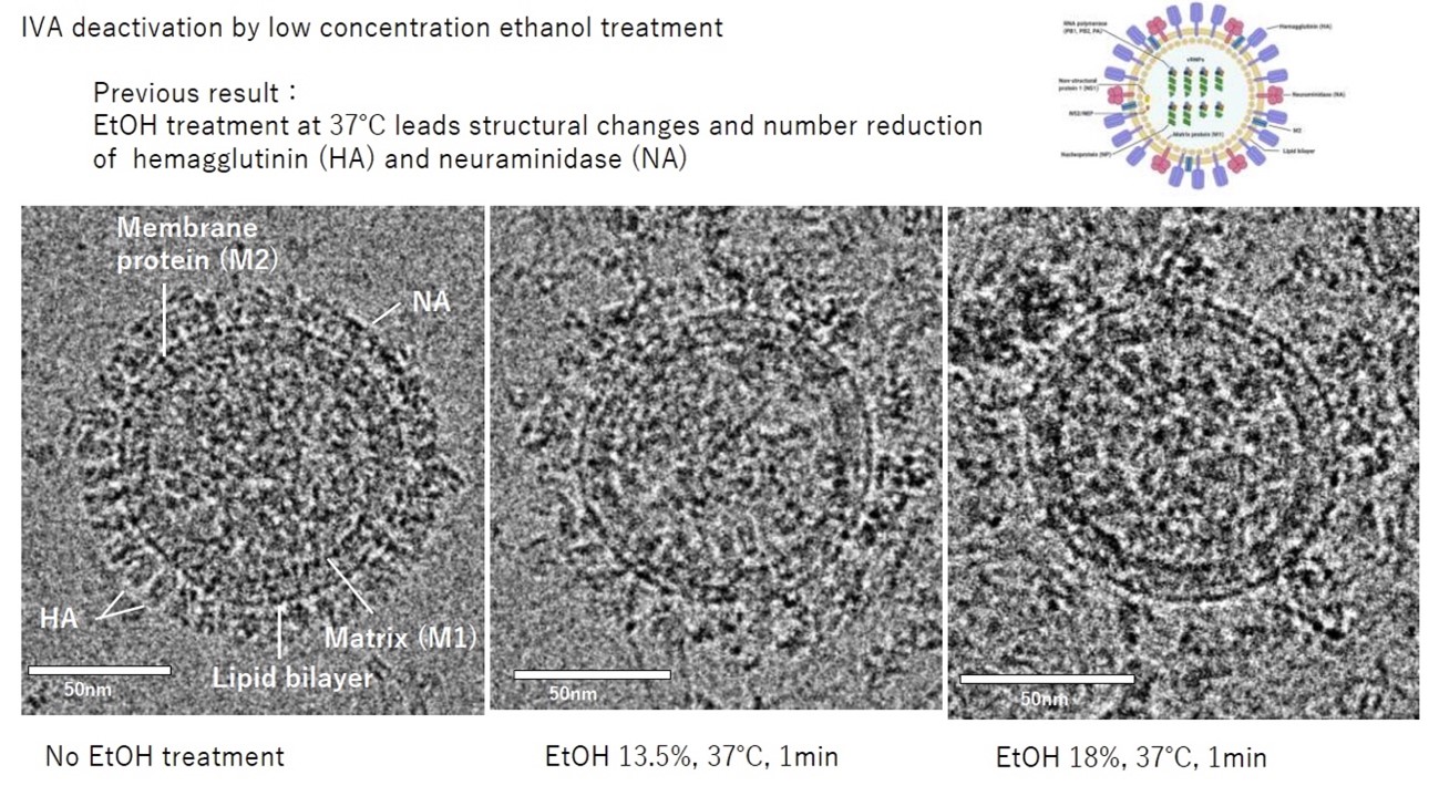
Figure 1. Conformational change on surface proteins on influenza-A virus after 1 minute exposure to EtOH
For further investigation of the ethanol effect, we develop a vapor diffusion device to control ethanol concentration in solution in a gentle manner. With using the device, which enables us to control the temperature and the concentration of EtOH carefully, we found that the deactivation and conformational change is dependent on both temperature and ethanol concentration.
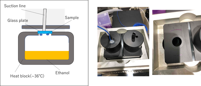
Figure 2. The vapor diffusion device (version 2). The ethanol vapor quickly adsorbs to the solution and creates an equilibrium with the target concentration
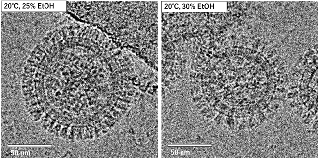
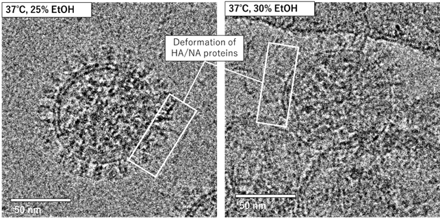
Figure 3. The cryo-TEM images of influenza-A virus after the treatment of EtOH by vapor diffusion method. The deformation of surface proteins were found when treated with EtOH at 37℃.
Dr.Takeshi Mise
(3) We aim to identify the lung condition of mice infected with the influenza A virus and the effect of ethanol vapor inhalation using micro-CT. This year, we observed the lungs of an uninfected mouse and established a micro-CT analysis method.

The micro-CT image of lungs from an uninfected mouse. A: Model drawing of lungs (blue) and mouse skeleton. B: The bronchi and alveoli of a mouse. C: Section image (0.7 mm thick) of Fig. A in the transverse plane. D: Section image (0.7 mm thick) of Fig. A in the longitudinal plane. E: Enlarged view of the yellow box in Fig. C. F: Enlarged view of the yellow box in Fig. D. H: Heart, Rl: Right lung, Ll: Left lung, V: Vessel, R: Rib, B: Bronchi, Ve: Vertebra, Br: Bronchiole, A: Alveolar sac.
Seita Taba
(4) Effect of temperature on EtOH resistance of influenza viruses The concentration required for inactivation of influenza viruses suspended in EtOH at room temperature (24°C) and at body temperature (37°C) was examined, and it was confirmed that at room temperature, more than 30% EtOH was required, while at 37°C, the virus was inactivated at a lower level near 20%.
Ethanol vapor treatment of influenza-infected mice
Based on the above results, we tested the therapeutic effect of introducing EtOH vapor to influenza-infected mice in collaboration with the Ishikawa Unit. We fabricated an ethanol induction device for mice (Fig. 1) based on a commercially available humidifier and introduced EtOH mist into the mice. We adjusted the concentration of ethanol supplied to the mice and examined the conditions under which the introduction of EtOH into mice would not have any adverse effects on the organism. EtOH was introduced into mice from the day before influenza infection. Results: The survival rate of EtOH-treated mice was clearly higher than that of non-treated mice.
Furthermore, a new EtOH inhaler (Fig. 2)is under construction for the purpose of introducing a higher concentration of EtOH vapor than the conventional ultrasonic mist system. The new device aims to further improve the therapeutic effect by heating the EtOH in the device to the target temperature and evaporating it to generate highly concentrated EtOH vapor, which is then introduced into the mouse.
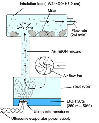
Fig. 1 Conceptual diagram of the EtOH mist induction system for mice The EtOH mist generated by the ultrasonic atomizer in the lower part is supplied to the inhalation box containing mice in the upper part by an air fan
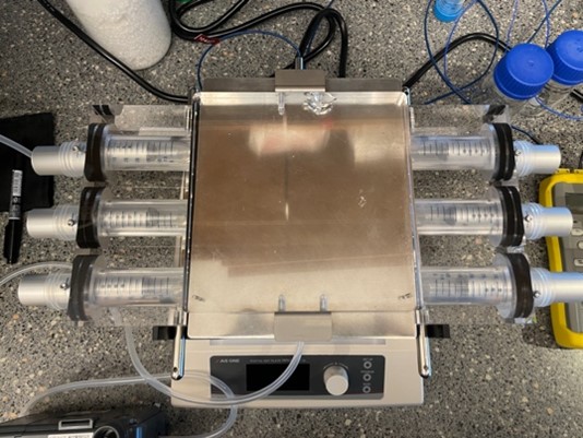
Fig. 2 New EtOH induction device
3.3 Wave energy project group (WEC Project)
OIST Wave Energy Project has been carrying out R&Ds to harness electricity from the moving water in the breaking wave zone since 2018, supported by Kokyo Tatemono Co. Ltd. in Tokyo, through their donation and contracted research.
Seragaki Site
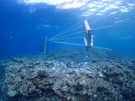
First YAMAHA MOTOR + OIST Wave Energy Converter has been installed at 3 m deep drop-off in front of the OIST Marine Science Station in April 2022. This small size Wave Energy Converter is suitable for remote island countries to harness electricity from perpetual ocean waves.
Ver.1
After installation in April, the turbine was updated in May (Ver.1) and data acquisition continued.
Ver.2
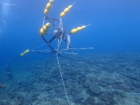
In September, Typhoon No. 11 caused rough sea conditions with wave heights exceeding 8M off Seragaki Fishing Port. It was a perfect opportunity for data acquisition, however, abnormal values were showed in the logger data. Waiting for sea conditions to improve, we examined the generator in the sea floor and found that the structure had been ripped away from the support trestle, and the cable had broken and fallen down. That seemed like significant damage but could be repaired and subsequently, the parts used in the holding section of the structure were replaced with aluminum parts to increase strength. After adjusting balance of the buoyancy structure, the generator was re-installed offshore of Seragaki Fishing Port. (Ver.2)
Ver.3
After the reinstallation was completed in November and about a month passed, the observation data from the generator was lost. We figured out the structure damage again. The solid aluminum parts had been bent like candy cane. Due to the above circumstances, the structure was carefully re-examined and modified to be as simple as possible (Ver.3) in March. We keep an eye on the structure condition and expect to receive efficient observation data during coming typhoon season.
Maldives
The Ducted WEC installation in Kandooma, Maldives had been suspended since 2021 and the generator and other equipment had remained on site. Consequently, the condition of those deteriorated significantly. Considering total cost to repair them and also reviewing the installation method, our decision was made to remove the generator and equipment once. In March 2023, the removal work was completed and Ducted WEC project was contractually terminated. When we develop advanced generator and find effective measure of installation, the project may recommence in Maldives.
4. Publications
4.1 Journals
- M. Tamai, S. Taba, T. Mise, M. Yamashita, H. Ishikawa, and T. Shintake, Effect of ethanol vapor inhalation treatment on lethal respiratory viral infection with Influenza A. The Journal of Infectious Diseases.doi: 10.1093/infdis/jiad089. Online AoP (2023)
- M. Tokoro Schreiber, C. Cassidy, M. Saidani, M. Wolf, A new electromagnetic lensing principle using the Aharonov-Bohm effect,arXiv (reviewer feedback received, update in progress), https://doi.org/10.48550/arXiv.2301.09980 (2023)
- K. Tani, R. Kanno, X.C. Ji, I. Satoh, Y. Kobayashi, M. Hall, L.J. Yu, Y. Kimura, A. Mizoguchi, B.M. Humbel, M.T. Madigan, Z.Y.Wang-Otomo, Rhodobacter capsulatus forms a compact crescent-shaped LH1-RC photocomplex, Nature Communications 14(1), 846. (2023)
- C. Cassidy, M. Tokoro Schreiber, M. Beleggia, J. Yamasaki, H. Adaniya, T. Shintake, Interpretation of mean free path values derived from off-axis electron holography amplitude measurements, Micron (2022) 162, 103346. doi:10.1016/j.micron.2022.103346
- H. Adaniya, M. Cheung, M. Yamashita, S. Taba, C. Cassidy, T. Shintake, Low energy scanning transmission electron microscopy applied to ice-embedded biological macromolecules, Microscopy (2022) dfac056.doi.org/10.1093/jmicro/dfac056
- A. Dhar, L.D.C. Jaubert, C. Cassidy, T. Shintake, N. Shannon,Observing Magnetic Monopoles in Spin Ice using Electron Holography, arXiv, https://doi.org/10.48550/arXiv.2112.01362 (2022)
- K. Tani, R. Kanno, K. Kurosawa, S. Takaichi, K.V P Nagashima, M. Hall, L.J. Yu, Y. Kimura, M.T. Madigan, A. Mizoguchi, B.M. Humbel, Z.Y. Wang-Otomo, An LH1-RC photocomplex from an extremophilic phototroph provides insight into origins of two photosynthesis proteins,Communications Biology 5(1), 1197. (2022)
- K. Tani, R. Kanno, R. Kikuchi, S. Kawamura, K. Nagashima, M. Hall, A. Takahashi, L. J. Yu, Y. Kimura, M. Madigan, A. Mizoguchi, B.M. Humbel, Z.Y. Wang-Otomo, Asymmetric Structure of the Native Rhodobacter sphaeroides Dimeric LH1-RC Complex, Nature Communications 13(1), 1904. (2022)
- K. Tani, K. Kobayashi, N. Hosogi, X.C. Ji, S. Nagashima, K.V.P. Nagashima, A. Izumida, K. Inoue, Y. Tsukatani, R. Kanno, M. Hall, A. Takahashi, L.J. Yu, I. Ishikawa, Y. Okura, M.T. Madigan, A. Mizoguchi, B.M. Humbel, Y. Kimura, Z.Y. Wang-Otomo, A Ca2+-Binding Motif Underlies the Unusual Properties of Certain Photosynthetic Bacterial Core Light-Harvesting Complexes, Journal of Biological Chemistry 298(6), 101967. (2022)
4.2 Books and other one-time publications
新竹 積「タンパク質結晶構造解析向け電子顕微鏡の開発」難波啓一他監修「クライオ電子顕微鏡ハンドブック」、第 7 章第 4 節、2023年1月20日発刊、株式会社エヌティーエス
4.3 Oral and Poster Presentations
- Tsumoru Shintake,Proposal of plane-parallel resonator configuration for high-NA EUV lithography, SPIE Photomask Technology + EUV Lithography, Septemver 2022, Monterey, California, United States
- 新竹 積、「熱燗の湯気で新型コロナや風邪を予防できる?」第2回 三鷹ネットワーク大学・オンライン、2023年6月18日
- Tsumoru Shintake,Can we block the center of an objective lens? From crystallography to telecentric microscopy, Photon Science Seminar at Stanford University, September 2022, United States
- Ryo Kanno, Oran Presentation.Old and New Methods for Cryo-EM・データ解析ハンズオンセミナ, 2022年10月26日-28日, 東北大学クライオ電子顕微鏡コース(INGEM)
5. Intellectual Property Rights and Other Specific Achievements
Nothing to report
6. Meetings and Events
Nothing to report
7. Other
7.1 Visitors
- 30 Jan, 2023 : Provost Michael Bruno, University of Hawaiʻi at Mānoa
- 15 May, 2022 : Delegation from Cabinet Office, Prime Minster Kishida
- 13 May, 2022 : Mr.Takeshi Furuichi, Chair of Kansai Keizai Doyukai
7.2 Media
Nothing to report


