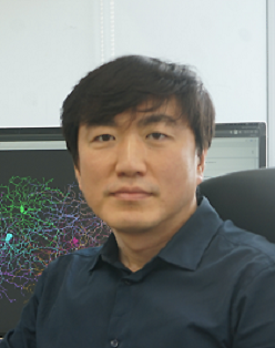
[Seminar] "Connectomic reconstruction of mouse cerebellar molecular layer from serial electron microscope images" by Dr. Jinseop Kim

Date
Location
Description
Abstract:
The regular organization of cerebellar cortical microcircuit is commonly accepted as well-understood, however, there remain a broad range of unexplored knowledges such as the ultrastructure of cells, subcellular wiring specificity, and single-cell variability. We aim to study the anatomy of cerebellar molecular layer through 3D reconstruction of densely-stained serial electron microscope images by multiple steps of image analysis. I will present our ongoing efforts towards the goal and a few preliminary discoveries. The reconstruction is performed through a semi-automated computer pipeline, whose components include an image alignment algorithm, an artificial intelligence for image segmentation, a user interface software for human validation of the segmentation, and background softwares for the management of validation process. Further analysis technologies to quantitatively describe the structure of cells and circuits are under development. The findings along with others include the facts regarding the spine distribution on Purkinje cells, synapses of granule cell ascending axon, and reciprocal connections between the inhibitory interneurons and can evoke open discussions on their biological implication.
Subscribe to the OIST Calendar: Right-click to download, then open in your calendar application.


