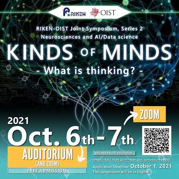
Kinds of Minds - what is thinking? 2nd OIST-RIKEN Symposium-

Date
Location
Description
Program
Click here to download
Applications
Apply Poster Poster presentation Deadline: September 27, 2021
Submit Poster If you have already applied for poster presentation and would like to submit your poster, please submit from here.
Registration Registration for participation Deadline: October 1, 2021
Symposium Abstract
-Message from Program Committee-
The last decade has seen strings of breakthroughs in both neuroscience and artificial intelligence research. While there have long been fruitful exchanges between these fields, there is increased recent interest in the borderlands, using computational ideas and techniques to inform biology, and vice versa. This symposium brings together researchers working on a diversity of topics circling around questions related to the mind. We hope to catalyze new ideas, future collaborations, and an appreciation for the diversity of views on the topic. Please join us.
Main Speakers and Abstracts
※Please find detailed schedule here
Session I-1 "Open systems approaches in neuroscience
- How can ideas from complexity science, dynamical systems, and embodied cognition help us understand the brain/mind?"
- Chaired by Dr. Kazuhiro SAKURADA
| 1 |
Dr. Andrea BENUCCI, Team Leader |
Stability of visual perception as a classification invariance in convolutional neuronal networks Our ability to perceive a stable visual world in the presence of continuous movements has puzzled researchers in the neuroscience field for a long time. We reformulated this problem in the context of hierarchical convolutional neural networks (CNN)—observed in the mammalian visual system—and show that perceptual stability can emerge from an optimization process that identifies image-defining features for accurate image classification in the presence of movements. Movement signals, multiplexed with visual inputs along overlapping convolutional layers, enabled movement-related classification invariance that, as a secondary process, permitted coordinate transformations from retinocentric to craniocentric reference frames. Classification invariance was reflected in clustered activity patterns in low dimensional manifolds associated with image categories and emerging in late CNN layers. Furthermore, movement signals affected visual responses according to modulatory principles observed experimentally during saccadic eye movements. Our findings provide a computational framework that unifies a multitude of biological observations on perceptual stability under optimality principles for image classification in artificial neural networks. |
|
| 2 |
Dr. Tom FROESE, Assistant Professor |
Exploring embodied social cognition with AI and neuroscience The social brain hypothesis has long been a dominant framework for understanding the relative increase of brain size in human evolution. However, recent large-scale analyses of brain-body data question its validity and generality as a principle of brain evolution. At the same time, theoretical advances in embodied social cognition have started to cast doubt on that framework’s adoption of methodological individualism as a starting point for thinking about brain function. In this talk, I will present agent-based models that permit us to systematically evaluate what real-time social interaction does to the complexity of neural activity, which holds a few surprises in store. And I will present preliminary data from our EEG hyperscanning lab, which confirms that social interaction indeed deeply affects brain functions. |
|
| 3 |
Dr. Kazuhisa SHIBATA, Team Leader |
Reading and manipulating information in the brain This talk will summarize technological developments in the field of human cognitive neuroscience over the past 20 years. First, I will describe multivariate analyses of brain signals measured by noninvasive neuroimaging methods such as functional magnetic resonance imaging (fMRI) and electroencephalography. These multivariate analyses have allowed researchers to read out or decode specific information from patterns of the brain signals. Second, I will explain real-time neurofeedback methods in which brain signals are presented to participants in a real-time manner to induce neural plasticity that in turn leads to changes in behavior. Third, I will introduce decoded neurofeedback, a hybrid technology that combined multivariate analyses with real-time fMRI neurofeedback. It has been shown that decoded neurofeedback induces specific fMRI signal patterns in a local and targeted brain region and changes specific behavior without participants’ awareness of the purpose of the experiment. Collectively, recent technological development in human cognitive neuroscience have enabled reading and manipulating information in the brain and substantially advanced both basic and clinical research. |
Session I-2 "Open systems approaches in neuroscience
- How can ideas from complexity science, dynamical systems, and embodied cognition help us understand the brain/mind?"
- Chaired by Dr. Kazuhiro SAKURADA
| 1 |
Dr. Jun TANI, Professor |
Studies on cognitive neurorobotics using the framework of predictive coding and active inference The focus of my research has been to investigate how cognitive agents can develop structural representation and functions via iterative interaction with the world, exercising agency and learning from resultant perceptual experience. For this purpose, my team has investigated various models analogous to predictive coding and active inference frameworks. For the last two decades, we have applied these frameworks to develop cognitive constructs for robots. The current talk introduces a set of emergent phenomena which we found in the robotics experiments. These findings inform us of possible non-trivial cognitive mechanisms in the brains. |
|
| 2 |
Dr. Reiko MAZUKA, Team Leader |
What we can learn from how human infants learn language in real time? In language learning research, it is crucial to understand the precise nature of input. This is particularly significant in real-life language acquisition, since it is known that how adults talk to infants (Infant-directed-speech, IDS) differs substantially from adult-to-adult speech. Yet, compared to “the mechanism” side of learning -- the structure and the procedures of learning, the nature of input from which learning must occur has not received the equal level of scrutiny. In this talk, we will demonstrate the importance of accurate characterization of input by showing two examples in phonological learning, in which failing to take into account relevant factors led to inaccurate conclusions – duration based consonant distinction acquisition, and the speech rate in IDS. These examples highlights the importance of examining not only the target features but also other factors that are potentially relevant in the input. |
|
| 3 |
Dr. Kenji DOYA, Professor |
The duality of inference and control as a key to understanding canonical cortical circuits An intriguing question about the brain is why the entire neocortex shares a canonical six-layer architecture while its posterior and anterior halves are engaged in sensory processing and motor control, respectively. Here we consider the hypothesis that the sensory and motor cortical circuits implement the dual computations for Bayesian inference and optimal control, respectively. Based on the architecture of the canonical cortical circuit, we explore how different cortical neurons may represent variables and implement computations for inference and control. |
Session II "Advances in neuroscience
-What are the strengths and limitations of current approaches?"
- Chaired by Dr. Sam REITER
| 1 |
Dr. Bernd KUHN, Professor |
Ca+ activity maps of astrocytes tagged by axo-astrocytic Astrocytes exhibit localized Ca2+ microdomain (MD) activity thought to be actively involved in information processing in the brain. However, the functional organization of Ca2+ MDs in |
|
| 2 |
Dr. Jun NAGAI, Team Leader |
Behind the scenes in neural circuits: Astrocyte regulation of behavior in health and disease Astrocytes, a type of non-neuronal glial cells, tile the entire central nervous system. I will report the latest insights on astrocyte signaling in the adult neural circuits, by using multiple integrated approaches, including calcium imaging, electrophysiology, opto/pharmaco-genetics, mouse behavioral tests, RNA-seq and new astrocyte manipulation tools that we recently developed. First, I will describe mechanisms of bi- directional neuron-astrocyte communications in the striatum that lead to hyperactivity and disrupted attention via a synaptogenic cue. Second, I will present how astrocytes respond to distinct perturbations and how we can use the molecular signaling information for phenotypic benefits in neurodegenerative disease mouse models, e.g. Huntington’s disease. Taken together, our findings show that signaling from astrocytes to neurons is sufficient per se to alter synapses, circuits and behavior. We also provide new tools to study such astrocyte-neuron dynamics. |
|
| 3 |
Dr. Yoko YAZAKI-SUGIYAMA, Associate Professor |
Neural Circuit for Social Authentication in Zebra Finch Song Learning Social interactions are essential when learning to communicate. In human speech and bird song, infants must acquire accurate vocalization patterns and learn to associate them with live tutors and not mimetic sources. However, the neural mechanism of social reality during vocal learning remains unknown. Here, we characterize a neural circuit for social authentication in support of accurate song learning in the zebra finch. We recorded neural activity in the noradrenergic command center, the locus coeruleus (LC), of juvenile birds during song learning from a live adult tutor. LC activity increased with real, not artificial, social information during learning that enhanced the precision and robustness of the learned song. During live social song learning, LC firing reconfigured long-term song-selective neural responsiveness in an auditory memory region, the caudomedial nidopallium (NCM). In accord, optogenetic inhibition of LC presynaptic signaling in the NCM disordered NCM neuronal responsiveness to live tutor singing and impaired song learning. These results demonstrate that the LC-NCM neural circuit integrates sensory evidence of real social interactions, distinct from song prosody, to authenticate song learning. The findings suggest a general mechanism for validating social information in brain development. |
|
| 4 |
Dr. Asuka TAKEISHI, Team Leader |
RIKEN CBS, Neural circuit of multisensory integration RIKEN Hakubi Research Team |
Neural mechanism of behavior modulation in C. elegans Animals make behavior decisions by integrating information of environmental stimuli and internal state. C. elegans, worms, has contributed greatly to elucidate the neural and molecular mechanisms of behavior decision with its advantages of the transparent body for fluorescent imaging, simple nervous system with complete connectome, and various genetic tools. Temperature is one of the most important environmental cues for animal survival, and worms thus can detect and respond to the subtle environmental temperature change. We have been studying how worms integrate sensory cues to modulate their temperature dependent behavior (thermotaxis) in order to understand the molecular and the neural basis of behavior decision. Here, I would like to present our recent findings on the mechanisms of starvation-dependent alteration of thermotaxis. Also, I would like to introduce our ongoing research to dissect the integration mechanisms of temperature and odor information, together with the newly developed techniques that we would like to incorporate in our future studies. |
Session III "AI/data science and brain/mind
-How can each inform the other?"
- Chaired by Dr. Yoko YAZAKI-SUGIYAMA
| 1 |
Dr. Tomomi SHIMOGORI, Team Leader |
RIKEN CBS, Laboratory for Molecular Mechanisms of Brain Development |
Cellular-resolution gene expression profiling in the neonatal marmoset brain reveals dynamic species- and region-specific differences Precise spatiotemporal control of gene expression in the developing brain is critical for neural circuit formation, and comprehensive expression mapping in the developing primate brain is crucial to understand brain function in health and disease. Here, we developed an unbiased, automated, large-scale, cellular-resolution in situ hybridization (ISH)-based gene expression profiling system (GePS) and companion analysis to reveal gene expression patterns in the neonatal New World marmoset cortex, thalamus, and striatum that are distinct from those in mice. Gene-ontology analysis of marmoset-specific genes revealed associations with catalytic activity in the visual cortex and neuropsychiatric disorders in the thalamus. Cortically expressed genes with clear area boundaries were used in a three- dimensional cortical surface mapping algorithm to delineate higher-order cortical areas not evident in two-dimensional ISH data. GePS provides a powerful platform to elucidate the molecular mechanisms underlying primate neurobiology and developmental psychiatric and neurological disorders. |
| 2 |
Dr. Sam REITER, Assistant Professor |
Exploring 2-stage sleep in octopus Human sleep can be divided into two stages, rapid eye movement (REM) and slow wave (SW) sleep, each with distinct behavioral and neural correlates as well as proposed functions. Two-stage sleep has been shown to be present in other mammals, as well as birds, reptiles, and fish. This suggests that it evolved very early in vertebrate evolution, has been maintained over the hundreds of millions of years separating these diverse animal groups, and therefore is of fundamental importance functionally. We recently found that octopuses, which evolved large brains and complex behaviors independently of the vertebrate lineage, also possess two stages of sleep (‘active’ and ‘inactive’). Each stage has a range of behavioral correlates resembling vertebrate REM and SW sleep. Octopus active sleep is characterized by the rapid transitioning through a series of brain-controlled skin patterns. Through high-resolution filming and computational analysis, we are attempting to relate octopus waking and sleeping skin patterns, and thus decode the evolving contents of octopus sleep. The possibility that two similar stages of sleep evolved convergently suggests a comparative approach may reveal general principles of sleep function. |
|
| 3 |
Dr. Henrik SKIBBE, Unit Leader |
Brain image analysis The goal of our unit is to develop algorithms and tools for the processing and analysis of multi-modal biomedical imaging data. Since we are a member of the Brain/MINDS project, much of our data are images of the brains of marmoset monkeys. First, I will introduce our ongoing work on processing neural tracer and gene expression images. One challenge in working with biomedical image data is that we must deal with uncertainty. As examples, I will present two methods we have developed in our unit: (1) A probabilistic axon tracking algorithm for large microscopy images. (2) A probabilistic convolutional neural network for predicting the evolution of white matter hyperintensities from MR images of the (human) brain. |
|
| 4 |
Dr. Tomoki FUKAI, Professor |
What can neural network models tell about brain activity: two separate strata in the cortex? Cell assembly refers to a group of neurons that are repeatedly coactivated to serve as a functional unit of cortical computation. The existence of such assemblies has often been indicated by the precise spatiotemporal firing patterns of a group of neurons as well as the clustered wiring patterns of cortical neurons. While machine learning offers various tools for detecting cell assemblies, here we show a different approach: we use a brain-inspired network model for segmenting cell- assembly structures in large-scale neural recording data. As a successful example, we report the assemblies of highly sparse firing (<1 Hz) neurons that are repeatedly activated across the superficial and deep layers of the rat motor cortex in a behavior-relevant manner. This study was also motivated by recent observations suggesting that local cortical circuits in the superficial layers and deep layers are, as a default, independent strata of brain computing. If so, the question is "who connects these strata, when and why?" Although our results cannot directly answer this question, they give an insight into the dynamics of cortical microcircuits in relation to this question. |
Poster Presenters
| Authors | Title | Poster | Theme | |
|---|---|---|---|---|
| 1 | Muhammad Febrian Rachmadi Brain Image Analysis Unit, CBS, RIKEN |
Probabilistic Deep Learning with Adversarial Training and Volume Interval Estimation - Better Ways to Perform and Evaluate Predictive Models for White Matter Hyperintensities Evolution | 2 | |
| 2 | Charissa Poon Brain Image Analysis Unit, CBS, RIKEN |
Deep learning applications for automated microscopy image analyses in the neurosciences | 3 | |
| 3 | Tomasz Maciej Rutkowski Cognitive Behavioral Assistive Technology Team, AIP, RIKEN |
AI Neurotechnology Application to Early Dementia Onset Elucidation from EEG in Visual Emotional and Reminiscent Interior Sorting Tasks | 1 | |
| 4 | Ni Lei Plant Genome Evolution Research Team, RNC, RIKEN |
Massive genome sequencing analysis of accelerated carbon-ion-induced mutations in Saccharomyces cerevisiae | 2 | |
| 5 | Lisa Okamoto Lab for Integrated Cellular Systems, IMS, RIKEN |
Meta-analysis of transcriptional regulatory networks for lipid metabolism in the neural cells from schizophrenia patients | 1 | |
| 6 | Momoe Sukegawa Lab for Neuroepitranscriptomics, BDR, RIKEN |
Investigating the behavioral impact of environmental factors on BALB/c strain mice | 1 | |
| 7 | Enrico Rinaldi iTHEMS, RIKEN |
Simulation-based inference for multi-type cortical circuits | 2 | |
| 8 | Lin Gu Machine Intelligence for Medical Engineering Team, AIP, RIKEN |
Enhance AI with Artificial Memory Inspired by Neuroscience | 2 | |
| 9 | Hiroshige Takeichi Open Systems Information Science Team, Information R&D and Strategy Headquarters, RIKEN |
Complex constraints in visual perception | 3 | |
| 10 | Roman Koshkin Neural Coding and Brain Computing Unit, OIST |
Role of Spontaneous Neural Activity in Memory Function | Poster | 2 |
| 11 | Shotaro Funai Physics and Biology Unit, OIST |
Comparison of neural activity for appreciation of Japanese tanka in human brain and artificial intelligence | Poster | 1 |
| 12 | Chen Lam Loh Embodied Cognitive Science Unit, OIST |
A Minimal Model for Interbrain Synchrony | Poster | 3 |
| 13 | Susana Ramírez Vizcaya Embodied Cognitive Science Unit, OIST |
Agents of Habit: Refining the Artificial Life Route to Artificial Intelligence | Poster | 3 |
| 14 | Toshitake Asabuki Neural Coding and Brain Computing Unit, OIST |
Learning spontaneously reactivatable prior distributions for causal inference | Poster | 2 |
| 15 | Zacharie Taoufiq Cellular and Molecular Synaptic Function Unit, OIST |
Molecular anatomy of brain synapses from living psychiatric patients | Poster | 1 |
| 16 | Ivan Shpurov Embodied Cognitive Science Unit, OIST |
Combining Self-critical dynamics and Hebbian learning to explain utility of bursty dynamics in neural networks | Poster | 3 |
| 17 | Sergio Verduzco Flores Neural Computation Unit, OIST |
Spinal cord plasticity "solves" the sensorimotor loop | Poster | 1 |
| 18 | Laura Mojica Embodied Cognitive Science Unit, OIST |
One body, four types of mind | Poster | 3 |
Attachments
Subscribe to the OIST Calendar: Right-click to download, then open in your calendar application.


