FY2018 Annual Report
Optical Neuroimaging Unit
Associate Professor Bernd Kuhn
Claudia Cecchetto, Miles Desforges;
Isabel Anai Echeverria Oviedo, Shinobu Nomura, Eltabbal Mohamed Mostafa Kamal;
Lina Mohamed Koronfel, Hiroko Chinone, Kazuo Mori, Leonidas Georgiou;
Soumen Jana, Bernd Kuhn, Neil Dalphin
Photo by Emiko Asato
Abstract
In the FY 2018 the Optical Neuroimaging Unit continued the projects of the previous years. Research projects focused on imaging neuronal activity in the brain of awake mice.
1. Staff
- Dr. Bernd Kuhn, Associate Professor
- Dr. Christopher J. Roome, Staff Scientist
- Dr. Sigita Augustinaite, Staff Scientist
- Dr. Shinobu Nomura, Postdoc
- Ray X. Lee, Ph.D. student
- Neil Dalphin, Ph.D. student
- Leonidas Georgiou, Ph.D. student
- Soumen Jana, Ph.D. student
- Lina Koronfel, Ph.D. student
- Dr. Kazuo Mori, Technical Assistant
- Hiroko Chinone, Research Unit Administrator
2. Collaborations
2.1 Post-traumatic behavioral development in adult mice
- Description: Behavioral study with fine scale analysis of post-traumatic behavioral development in adult mice
- Type of collaboration: Joint research
- Researchers:
- Associate Professor Greg Stevens, OIST and Vrije Universiteit Amsterdam
2.2 Imaging cortical activity during spontaneous behavior
- Description: Calcium imaging of cortical neurons in different regions and layers during spontaneous behavior
- Type of collaboration: Joint research
- Researchers:
- Associate Professor Greg Stevens, OIST and Vrije Universiteit Amsterdam
2.3 High-resolution imaging of the barrel cortex through VSD and LFP recordings
- Description: Combining high-resolution electical recording with electrode arrays and voltage imaging
- Type of collaboration: Joint research
- Researchers:
- Associate Professor Stefano Vassanelli, Padua University, Italy
2.4 Imaging average membrane potential with ANNINE-6 in layer 1 of barrel cortex of the mouse
- Description: Imaging voltage from cortical layer 1 in mice; synthesis of new voltage-sensitive dyes
- Type of collaboration: Joint research
- Researchers:
- Dr. Eugene Khaskin, Science and Technology Group, OIST
3. Activities and Findings
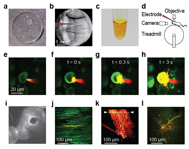
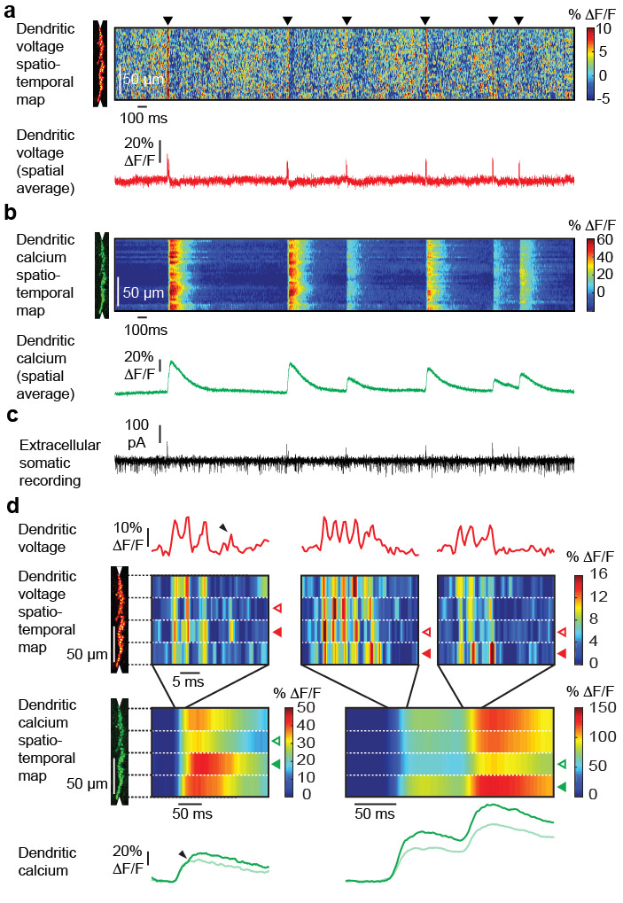
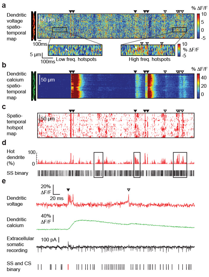
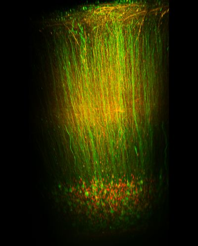
3.1 Simultaneous dendritic voltage and calcium imaging and somatic recording from Purkinje neurons in awake mice
Spatio-temporal maps of dendritic signalling and their relationship with somatic output is fundamental to neuronal information processing, yet remain unexplored in awake animals. Here, we combine simultaneous sub-millisecond voltage and calcium two-photon imaging from distal spiny dendrites, with somatic electrical recording from spontaneously active cerebellar Purkinje neurons (PN) in awake mice. We detect discrete 1-2 ms supra-threshold voltage spikelets in the distal spiny dendrites during dendritic complex spikes. Spikelets and their calcium correlates are highly heterogeneous in number, timing and spatial distribution within and between complex spikes. Back-propagating simple spikes are highly attenuated. Highly variable 5-10 ms voltage hotspots are localized to fine dendritic processes and are reduced in size and frequency by lidocaine and CNQX. Hotspots correlated with somatic output but also, at high frequency, trigger purely dendritic calcium spikes. Summarizing, spatio-temporal signalling in PNs is far more complex, dynamic, and fine scaled than anticipated, even in resting animals.
3.2 Imaging average membrane potential with ANNINE-6 in layer 1 of barrel cortex of the mouse
Membrane voltage oscillations in layer 1 of primary sensory cortices might be important indicators of cortical gain control, attentional focusing, and signal integration. However, electric field recordings are hampered by the low seal resistance of electrodes close to the brain surface. To study layer 1 membrane voltage oscillations, we synthesized a new voltage-sensitive dye, di1-ANNINE-6plus, that can diffuse into tissue. We applied it with a new surgery, leaving the dura intact but allowing injection of large quantities of staining solution, and imaged cortical membrane potential oscillations with two-photon microscopy depth-resolved (25 to 100 µm below dura) in anesthetized and awake mice. We found delta (0.5-4 Hz), theta (4-10 Hz), low beta (10-20 Hz), and low gamma (30-40 Hz) oscillations. All oscillations were stronger in awake animals. While the power of delta, theta, and low beta oscillations increased with depth, the power of low gamma was more constant throughout layer 1. These findings identify layer 1 as an important coordination hub for the dynamic binding process of neurons mediated by oscillations.
3.3 Two-photon imaging of layer 6 corticothalamic feedback in a behaving mouse
Layer 6 (L6), the deepest lamina of cerebral cortex, is one of the key structures regulating behavior state related information processing within cortex and various subcortical areas. However, very little is known about the functional significance of different L6 circuits in vivo. In this project, we focus on primary visual cortex L6 feedback projections to visual thalamus (dorsal lateral geniculate nucleus, dLGN) which regulate visual signal transmission from retina to cortex. We perform calcium imaging of retrogradely marked L6 corticothalamic (CT) neurons with 2P microscopy in a head-fixed Ntsr1-cre mouse. We record neuronal activity from the same neurons for several hours and / or repeat during different days while presenting visual stimuli and monitoring the activity of a mouse. In this way, we can study the corticothalamic feedback during different behavior states, ranging from full alertness to sleep.
3.4 GRACE: hiGh-Resolution imAging of the barrel CortEx through VSD and LFP recordings
The aim of this research project is to develop an innovative and advanced dual approach to study the barrel cortex in mice, combining Voltage Sensitive Dye (VSD) imaging and high-resolution electrical recordings. The research will be carried out between Japan and Italy: at OIST (Okinawa, Japan) during the initial outgoing phase and at the Department of Biomedical Sciences of the University of Padova during the returning phase.
Sensory-evoked activity in the neocortex is known to manifest in the form of propagating waves but up to now there are no studies directed towards a high-resolution mapping of these waves in the barrel cortex in vivo. VSD imaging will be performed simultaneously with high-resolution electrical mapping of Local Field Potentials (LFPs) through CMOS-based implantable neural probes developed within an EU project coordinated by UNIPD (CyberRat; FP7, #216528). Once fully established in OIST, this dual method will be transferred to UNIPD and will allow the study of neuronal signal propagation through a 3D architecture in living tissue with simultaneous high-resolution optical and electrical recordings.
3.5 Imaging neuron glia interaction in awake mice
One of the most exciting modern hypothesis in neuroscience is that astrocytes respond to neuronal signals with fast (<500ms) calcium transients that in turn can influence neuronal activity. However, it is controversial whether astrocytes respond reliably to neuronal signals in vivo. We developed a method that allows us to simultaneously record the activity of cortical astrocytes and thalamocortical axons at high contrast in the awake mouse. The method exploits the poorly characterized anterograde trans-cellular transfer properties of adeno-associated viruses (AAVs). We found that AAVs can transfer via axons to both neurons and astrocytes. This property allows us to express genetically encoded calcium indicators in axons and sparse contacting astrocytes and image their interactions with two-photon microscopy through a cranial window. We are investigating how different behavioral states (i.e. whisker stimulation, running, resting, sleeping, stress) influence the communication between axons and astrocyte microdomains in the somatosensory cortex of mice. Our findings challenge the spatial specificity of AAVs and allow us to record astrocyte-neuron interactions at high contrast under physiological conditions.
3.6 Imaging PKA activity in vivo
In the CNS, protein kinase A (PKA) is controlled by neuromodulators through G-protein-coupled receptors, therefore it is thought to be involved in multiple brain functions. However, PKA activity has not been observed with cellular or subcellular resolution in vivo.
In this study we imaged PKA activity of cortical neurons in awake mice. A genetically encoded single GFP-based PKA sensor, GakdYmut, was expressed after adeno-associated viral gene transfer. Mice were head fixed on a cylindrical treadmill and were allowed to walk ad libitum during imaging using two-photon microscopy through a chronic cranial window.
In somatosensory cortex, PKA activity rose in dendrites and somata of neurons in layer II/III and V with the onset of locomotion, and then reached a maximal amplitude after walking offset. PKA activity in dendrites and somata peaked 20 ± 9 seconds (n = 685) and 20± 6 seconds (n = 372), respectively, after the offset of locomotion, with maximal amplitudes of ΔF/F = 14%. PKA activity from labeled dendrites (43%) and somata (22%) showed synchronous responses greater than 2.5% ΔF/F (20s time window), in the same field of view. Similar PKA activity patterns were detected in the posterior parietal cortex, but not in the anterior cingulate cortex. Simultaneous imaging of PKA and calcium with the red calcium indicator protein rGECO revealed that PKA activation is independent of calcium influx due to spiking or bursting. We will try to identify neuromodulators which affect locomotion-related PKA activity in cortical neurons using pharmacology and electrophysiology.
3.7 Social relationship and brain hemisphere-specific correlates explain stress incubation in adult mice
While stress reactions can emerge long after the triggering event, it remains elusive how they emerge after a protracted, seemingly stress-free period, during which stress incubates. Here, we study the behavioral development in mice isolated after observing an aggressive encounter inflicted upon their pair-housed partners and compared the results with those in multiple control paradigms. Mice isolated following the acute witnessing social stress gradually developed a wide range of long-term differences, compared to control, of their spontaneous behaviors, social interactions, and physiological conditions, including hemisphere-specific structural differences in multiple cerebrocortical areas. We developed a spatially resolved fine-scale behavioral analysis and applied it to standard behavioral tests. It reveals that the seemingly sudden emergent behavioral differences developed gradually. Interestingly, these behavioral differences were not observed if the aggressive encounter happened to a stranger mouse, suggesting that social relationship underlies the stress development in this paradigm, which may have important disease-relevant links.
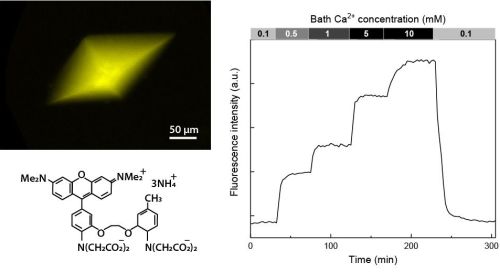
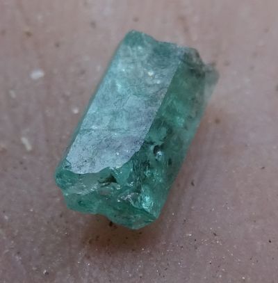
3.8 Imaging Ca2+ concentration and pH in nanopores/channels of protein crystals
Protein crystals are nanoporous materials. Despite this important characteristic, little is known about the conditions in the pores, also called channels. Here, we describe a method to study the calcium concentration and pH in the nanopores of thaumatin and lysozyme crystals. We load the crystal nanopores with fluorescent indicators and then perfuse the crystals with solutions of different calcium concentrations and pH while reading out the crystal’s fluorescence intensity with confocal microscopy. By calibrating the fluorescence signal we can determine the calcium concentration and pH in the nanopores. For the pH in thaumatin nanopores measured with the ratiometric pH sensor SNARF-1 we find a -0.7 pH shift compared to the bath pH corresponding to a 5-fold higher proton concentration. This is similar to the -0.3 pH shift found in lysozyme nanopores. With single-wavelength probes we find that the calcium concentration in thaumatin crystal nanopores is the same as in the bath while it is 0.24 times lower in lysozyme nanopores. Summarizing, our experiments show that calcium concentration and pH in the nanopores of protein crystals can deviate significantly from that in the bath. In general, the described method can be applied for testing a wide range of ion or small molecule concentrations in transparent nanoporous materials not only with ratiometric but also with single wavelength fluorescent indicators.
4. Publications
4.1 Journals
-
C.J. Roome* and B. Kuhn* (2018) Simultaneous dendritic voltage and calcium imaging and somatic recording from Purkinje neurons in awake mice. Nature communications, DOI: 10.1038/s41467-018-05900-3
-
K. Mori* and B. Kuhn* (2018) Imaging Ca2+ concentration and pH in nanopores/channels of protein crystals. J.Phys.Chem.B 122, 42, 9646-9653
-
S. Augustinaite and P. Heggelund (2018) Short-term synaptic depression in the feedforward inhibitory circuit in the dorsal lateral geniculate nucleus. Neuroscience 384, 76-86.
4.2 Books and Other One-Time Publications
Nothing to report
4.3 Oral and Poster Presentations
- S. Augustinaite (2019) Two-photon imaging of the deepest cortical layer (layer 6) in the behaving mouse. Internal Seminar at OIST
- R.X. Lee and B. Kuhn (2018) Neuronal dynamic framework of cerebral cortical networks for spontaneous behaviors. ICIIBMS 2018, Bangkok, Thailand
- S. Augustinaite and B. Kuhn (2018) V1 layer 6 corticothalamic feedback encodes behavioral state by complementary activity of two neuronal populations. Resonance Biology for Innovative Bioimaging, Meeting of Bioimaging for Young Researchers, OIST
- C.J. Roome and B. Kuhn (2018) Simultaneous dendritic voltage and calcium imaging and somatic recording from Purkinje neurons in awake mice. Resonance Biology for Innovative Bioimaging, Meeting of Bioimaging for Young Researchers, OIST
- N. Dalphin and B. Kuhn (2018) Simultaneous voltage and calcium imaging of mouse barrel cortex, in vivo. Resonance Biology for Innovative Bioimaging, Meeting of Bioimaging for Young Researchers, OIST
- S. Nomura and B. Kuhn (2018) Imaging Protein Kinase A Activity in Somatosensory Cortex of Behaving Mice. Resonance Biology for Innovative Bioimaging, Meeting of Bioimaging for Young Researchers, OIST
- L. Georgiou and B. Kuhn (2018) AAV mediated trans-synaptic tagging of astrocytes: A novel tool for studying neuron-astrocyte interactions. Society for Neuroscience Annual Meeting, San Diego, CA, USA
- S. Augustinaite and B. Kuhn (2018) V1 layer 6 corticothalamic feedback encodes behavioral state by complementary activity of two neuronal populations. Society for Neuroscience Annual Meeting, San Diego, CA, USA
- S. Nomura and B. Kuhn (2018) Imaging cortical Protein Kinase A activity in dendrites and somata of awake mice. FENS 2018, Berlin, Germany.
- Eva Berlot, Noah Steinberg, Tyler Manning, and Lina Koronfel (2018) Analyzing the Effect of Noise Correlations on Decoding Neuronal Recording. Summer School in Computational Sensory-Motor Neuroscience, University of Minnesota, Minneapolis, MN, USA.
4.4 Invited Talks
- OIST Minisymposium The 16th International Membrane Research Forum, OIST. Bernd Kuhn: Membrane voltage imaging with the pure electrochromic probe ANNINE-6. Organized by Prof. A. Kusumi, K. Kono (OIST). March 20, 2019.
- 5th Core-to-Core International Symposium "3D Lab-Exchange Program”, OIST. Bernd Kuhn: Voltage imaging from single neurons in awake animals. Organized by Prof. T. Inoue (Waseda University). February 27, 2019.
- Photonics Workshop, OIST. Bernd Kuhn: Imaging neuronal activity with two-photon microscopy in awake mice. Organized by Prof. J. Hayase (KEIO University). November 30, 2018.
- Resonance Biology for Innovative Bioimaging, Meeting of Bioimaging for Young Researchers “Chanpuru”, OIST. Bernd Kuhn: Two-photon microscopy: Principles and in vivo applications. Organized by Drs. K. Otomo, H. Ishii (Hokkaido University). October 29, 2018.
- Merocyanine 540 - 45+1 Years of Voltage Imaging, Woods Hole, MA, USA. Bernd Kuhn: Simultaneous spatio-temporal dendritic voltage/calcium mapping and somatic recording from Purkinje neurons in awake mice. Organized by Profs. L.B. Cohen, D. Zecevic, B.M. Salzberg, August 30-September 1, 2018.
- OIST-KAIST Joint Symposium, OIST. Bernd Kuhn: Simultaneous spatio-temporal dendritic voltage/calcium mapping and somatic recording from Purkinje neurons in awake mice. Organized by Profs. A. Shen, B. Kuhn (OIST). August 20, 2018.
- Molecular Biology Department, Princeton University, Princeton, NJ, U.S.A. Bernd Kuhn: Adeno-Associated Virus (AAV) transfer from axons to astrocytes. Invited by Prof. S.J. Flint. June 16, 2018.
- Princeton Neuroscience Institute, Princeton University, Princeton, NJ, U.S.A. Bernd Kuhn: Simultaneous spatio-temporal dendritic voltage/calcium mapping and somatic recording from Purkinje neurons in awake mice. Invited by Prof. S.S.-H. Wang. June 15, 2018.
5. Intellectual Property Rights and Other Specific Achievements
Nothing to report
6. Meetings and Events
6.1 Workshop: Okinawa Computational Neuroscience Course 2018
- Date: June 25- July 12, 2018
- Venue: OIST Seaside House
- Organizers: Drs. E. De Schutter, K. Doya, J. Wickens, B. Kuhn
- Speaker (among others): Bernd Kuhn
- Title 1: Voltage-gated channels and the Hodgkin-Huxley model of neuronal activity
- Title 2: Functional Optical Imaging Methods
6.2 Onna-son/OIST Children’s School of Science 2018
- Date: August 20-24, 2018
- Venue: Fureai Taiken Center
- Speaker (among others): Bernd Kuhn
- Topic: Palaeontology of Okinawa for Grade 1-3
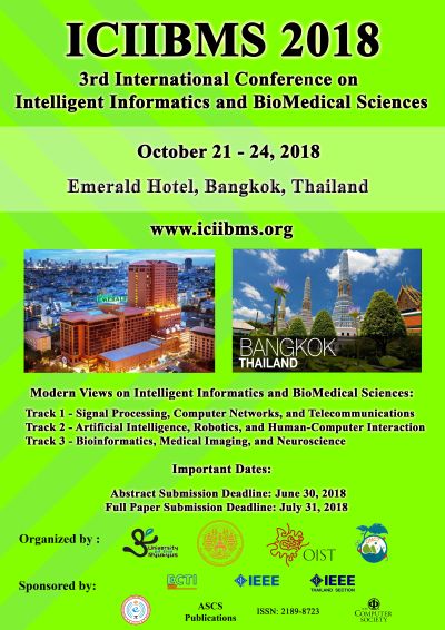
6.3 ICIIBMS 2018
- The Second International Conference on Intelligent Informatics and BioMedical Sciences (ICIIBMS) is in the planning phase.
- Date: October 21-24, 2018
- Venue: Bangkok
- Organized by researchers of the University of the Ryukyus, the Okinawa National College of Technology, and OIST
- 150 participants
6.4 Voltage Imaging Mini-symposium
- Date:
- Venue: OIST
- Speakers:
- Srjan Antic (UCONN)
- Dejan Zecevic (Yale University)
- Takashi Tominaga (Tokushima Bunri University)
- Sachiko Tsuda (Saitama University)
- Bradley Baker (KIST)
- Kenji Doya (OIST)
- Yuki Sudo (Okayama University)
- 'Larry' Laurance B. Cohen (Yale University, KIST)
- Yuki Bando (Hamamatsu University)
- Ryuichi Nakajima (KIST, SPEC corporation)
- Claudia Cecchetto (OIST)
- Chris Roome (OIST)
- Topic: Voltage Imaging
Kenji Doya, Bernd Kuhn, Laurance B. Cohen, Ryuichi Nakajima, Sachiko Tsuda, Yuki Sudo
7. Other
7.1 Planning of ICIIBMS 2019
- The Fourth International Conference on Intelligent Informatics and BioMedical Sciences (ICIIBMS) is in the planning phase.
- Date: November 21-24, 2019
- Venue: Shanghai, China
- Organized by researchers of the University of the Ryukyus, the Okinawa National College of Technology, and OIST