FY2017 Annual Report
Optical Neuroimaging Unit
Associate Professor Bernd Kuhn
Photo by Emiko Asato
Abstract
In the FY 2017 the Optical Neuroimaging Unit continued several imaging projects. Most research projects focused on imaging neuronal activity in the mouse brain.
1. Staff
- Dr. Bernd Kuhn, Associate Professor
- Dr. Christopher J. Roome, Postdoc
- Dr. Sigita Augustinaite, Postdoc
- Dr. Shinobu Nomura, Postdoc
- Ray X. Lee, Ph.D. student
- Neil Dalphin, Ph.D. student
- Leonidas Georgiou, Ph.D. student
- Soumen Jana, Ph.D. student
- Lina Koronfel, Ph.D. student
- Dr. Kazuo Mori, Technical Assistant
- Hiroko Chinone, Research Unit Administrator
2. Collaborations
2.1 Post-traumatic behavioral development in adult mice
- Description: Behavioral study with fine scale analysis of post-traumatic behavioral development in adult mice
- Type of collaboration: Joint research
- Researchers:
- Associate Professor Greg Stevens, OIST and Vrije Universiteit Amsterdam
2.2 Imaging cortical activity during spontaneous behavior
- Description: Calcium imaging of cortical neurons in different regions and layers during spontaneous behavior
- Type of collaboration: Joint research
- Researchers:
- Associate Professor Greg Stevens, OIST and Vrije Universiteit Amsterdam
3. Activities and Findings
3.1 Simultaneous dendritic voltage and calcium imaging and somatic recording from Purkinje neurons in awake mice
Spatio-temporal maps of dendritic signalling and their relationship with somatic output is fundamental to neuronal information processing, yet remain unexplored in awake animals. Here, we combine simultaneous sub-millisecond voltage and calcium two-photon imaging from distal spiny dendrites, with somatic electrical recording from spontaneously active cerebellar Purkinje neurons (PN) in awake mice. We detect discrete 1-2 ms supra-threshold voltage spikelets in the distal spiny dendrites during dendritic complex spikes. Spikelets and their calcium correlates are highly heterogeneous in number, timing and spatial distribution within and between complex spikes. Back-propagating simple spikes are highly attenuated. Highly variable 5-10 ms voltage hotspots are localized to fine dendritic processes and are reduced in size and frequency by lidocaine and CNQX. Hotspots correlated with somatic output but also, at high frequency, trigger purely dendritic calcium spikes. Summarizing, spatio-temporal signalling in PNs is far more complex, dynamic, and fine scaled than anticipated, even in resting animals.

Figure 1 A single Purkinje neuron was filled with ANNINE dye through a pipette at the cell body (top). A typical neuron has a cell body with many tree-shaped processes called dendrites, and a long cable-like process called the axon. The dendrites receive inputs, process information and pass it to the cell body. When a Purkinje neuron receives strong input, the entire dendrite gets activated (upstrokes in red trace) and the calcium ions flow into the neuron (transients in green trace). The voltage signals are 10-100 times faster than the variations in calcium levels.
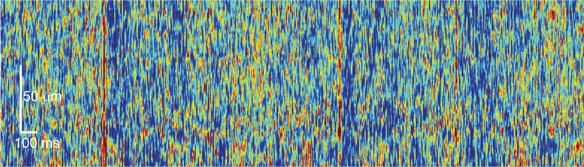
Figure 2 Spatially and temporally resolved map of a Purkinje neuron dendrite. Every red hotspot indicates an input to the dendrite. The red lines indicate activation of the full dendritic tree.
3.2 Imaging average membrane potential with ANNINE-6 in layer 1 of barrel cortex of the mouse
Layer 1 in somatosensory cortex receives large quantities of cortico-cortical axonal innervation on distal dendrites of layer 2/3 and layer 5 distal dendrites of pyramidal neurons, theorized to exert gain control in the context of attentional focusing. Despite its importance, layer 1 has been difficult to study, as electrodes cannot focus on it. Here, we use two-photon microscopy for simultaneous bulk voltage and subcellular calcium imaging in somatosensory cortex of the mouse to study sensory responses during a standard stimulation, and during an oddball experiment. The technique allows depth resolved voltage measurements of average membrane potentials and dendritic calcium transients.
3.3 2P imaging of layer 6 corticothalamic feedback in a behaving mouse
Layer 6 (L6), the deepest lamina of cerebral cortex, is one of the key structures regulating behavior state related information processing within cortex and various subcortical areas. However, very little is known about the functional significance of different L6 circuits in vivo. In this project, we focus on primary visual cortex L6 feedback projections to visual thalamus (dorsal lateral geniculate nucleus, dLGN) which regulate visual signal transmission from retina to cortex. We perform calcium imaging of retrogradely marked L6 corticothalamic (CT) neurons with 2P microscopy in a head-fixed Ntsr1-cre mouse. We record neuronal activity from the same neurons for several hours and / or repeat during different days while presenting visual stimuli and monitoring the activity of a mouse. In this way, we can study the corticothalamic feedback during different behavior states, ranging from full alertness to sleep.
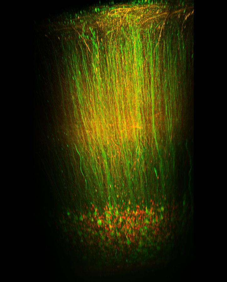
Figure 3 Reconstruction of Layer 6 neurons.
3.4 Imaging neuron glia interaction in awake mice
One of the most exciting modern hypothesis in neuroscience is that astrocytes respond to neuronal signals with fast (<500ms) calcium transients that in turn can influence neuronal activity. However, it is controversial whether astrocytes respond reliably to neuronal signals in vivo. We developed a method that allows us to simultaneously record the activity of cortical astrocytes and thalamocortical axons at high contrast in the awake mouse. The method exploits the poorly characterized anterograde trans-cellular transfer properties of adeno-associated viruses (AAVs). We found that AAVs can transfer via axons to both neurons and astrocytes. This property allows us to express genetically encoded calcium indicators in axons and sparse contacting astrocytes and image their interactions with two-photon microscopy through a cranial window. We are investigating how different behavioral states (i.e. whisker stimulation, running, resting, sleeping, stress) influence the communication between axons and astrocyte microdomains in the somatosensory cortex of mice. Our findings challenge the spatial specificity of AAVs and allow us to record astrocyte-neuron interactions at high contrast under physiological conditions.
3.5 Imaging PKA activity in vivo
Protein kinase A (PKA) is an important enzym in the signaling cascade connecting neuronal activity and expression and modulation of proteins. We use genetically encoded indicators to image PKA activity in vivo.
3.6 Uncover PTSD-like internal psychological development
Despite a broad research focus on the diverse symptoms of post-traumatic stress disorder (PTSD), the gradual emergence of symptoms following trauma exposure remains as a major conceptual challenge. I identified and examined behavioral development in mice isolated after observing a traumatic event inflicted upon their pair-housed partners. Compared to mice isolated without traumatic experience, traumatized mice displayed stractural changes in the forbrain. Fine-scale behavioral analysis showed that anxiety-related reactions gradually increased through isolation, while the early phases were independent of acute stress reactions. Instead of social avoidance, traumatized mice presented long-term social apathy to strangers. These behaviors were not observed if the traumatic event happened to strangers. Additionally, traumatized mice showed rebound responses to their partners after a long separation. Concluding, a single common factor, affectional bonding, underlies the rather independent development of PTSD-like behaviors in mice. The observed psychological homology between mice and humans emphasizes the potential of rodent models to untangle subtle psychobehavioral development in humans.
3.7 Toward neuronal dynamic framework for spontaneous behaviors
Despite as a well-known nature that animals outside from any behavioral task display irregular organization of spontaneous behaviors, the underlying neuronal and behavioral associations remains unclear. We approach this challenge by investigating neuronal population activities across cerebral cortical layers and areas using two-photon calcium imaging in head-restrained mice. The data characterized distinct dynamic features of neuronal population activity embedded in their region-specified static features. With the dominant representation of behavioral state, temporal encoding of behavioral dynamics varied along multiple aspects of a single population-level dynamical process. By testing predictive power for future behavioral and neuronal states, the results demonstrate an open-loop control for movement generation and termination and a close-loop control for ongoing movement. Concluding, two distinct control mechanisms of neurobehavioral association are employed in producing dynamic patterns of spontaneous behaviors.
3.8 Imaging Ca2+ concentration and pH in nanopores/channels of protein crystals
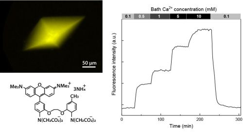
Protein crystals are nanoporous materials. Despite this important characteristic, little is known about the conditions in the pores, also called channels. Here, we describe a method to study the calcium concentration and pH in the nanopores of thaumatin and lysozyme crystals. We load the crystal nanopores with fluorescent indicators and then perfuse the crystals with solutions of different calcium concentrations and pH while reading out the crystal’s fluorescence intensity with confocal microscopy. By calibrating the fluorescence signal we can determine the calcium concentration and pH in the nanopores. For the pH in thaumatin nanopores measured with the ratiometric pH sensor SNARF-1 we find a -0.7 pH shift compared to the bath pH corresponding to a 5-fold higher proton concentration. This is similar to the -0.3 pH shift found in lysozyme nanopores. With single-wavelength probes we find that the calcium concentration in thaumatin crystal nanopores is the same as in the bath while it is 0.24 times lower in lysozyme nanopores. Summarizing, our experiments show that calcium concentration and pH in the nanopores of protein crystals can deviate significantly from that in the bath. In general, the described method can be applied for testing a wide range of ion or small molecule concentrations in transparent nanoporous materials not only with ratiometric but also with single wavelength fluorescent indicators.
4. Publications
4.1 Journals
Nothing to report
4.2 Books and other one-time publications
Nothing to report
4.3 Oral and Poster Presentations
-
S. Augustinaite, B. Kuhn (2017) 2-P imaging of visual cortex layer 6 corticothalamic feedback in the behaving mouse. Society for Neuroscience Annual Meeting, Washington, DC
-
S. Augustinaite, B. Kuhn (2017) 2-P imaging of visual cortex layer 6 corticothalamic feedback in the behaving mouse. The 40th Annual Meeting of the Japan Neuroscience Society, Chiba, Japan
-
R.X. Lee, G.J. Stephens, B. Kuhn (2017) Computational behavioral analysis of acute psychosocial trauma reveals gradually increasing stress reactions in adult mice. CNS – Organization for Computational Neurosciences, Antwerp, Belgium
-
L. Georgiou, B. Kuhn (2017) A novel method for labeling cortical astrocytes in vivo. 13th European Meeting on Glial Cells in Health and Disease, Edinburgh, GB
-
B. Kuhn (2017) Ins and outs of dendritic integration in awake mice. Invited by: Prof. W.-H. Yung, Sha Tin, Hong Kong, China
-
Sigita Augustinaite (2018) Complementary V1 layer 6 corticothalamic feedback encodes behavioral state. Seminar at Padua Neuroscience Center, University of Padua, Padua, Italy. Invited by Prof. Stefano Vassanelli.
-
B. Kuhn (2018) Simultaneous dendritic voltage and calcium imaging and somatic recording from Purkinje neurons in awake mice. Seminar at Padua Neuroscience Center, University of Padua, Padua, Italy. Invited by Prof. Stefano Vassanelli.
-
S. Augustinaite, B. Kuhn (2018) Complementary V1 layer 6 corticothalamic feedback encodes behavioral state. Gordon Research Conference “Thalamocortical Interactions: Development, Function and Dysfunction of Thalamocortical Networks”, Lucca (Barga), Italy.
-
S. Augustinaite (2018) Complementary V1 layer 6 corticothalamic feedback encodes behavioral state. Gordon Research Seminar “Thalamocortical Interactions: Exploring the Role of Thalamocortical Interactions in Sensory and Motor Networks and Development”, Lucca (Barga), Ital
5. Intellectual Property Rights and Other Specific Achievements
Nothing to report
6. Meetings and Events
6.1 Workshop: Okinawa Computational Neuroscience Course 2017
- Date: June 26- July 13, 2017
- Venue: OIST Seaside House
- Organizers: Drs. E. De Schutter, K. Doya, J. Wickens, B. Kuhn
- Speaker (among others): Bernd Kuhn
- Title 1: Voltage-gated channels and the Hodgkin-Huxley model of neuronal activity
- Title 2: Functional Optical Imaging Methods
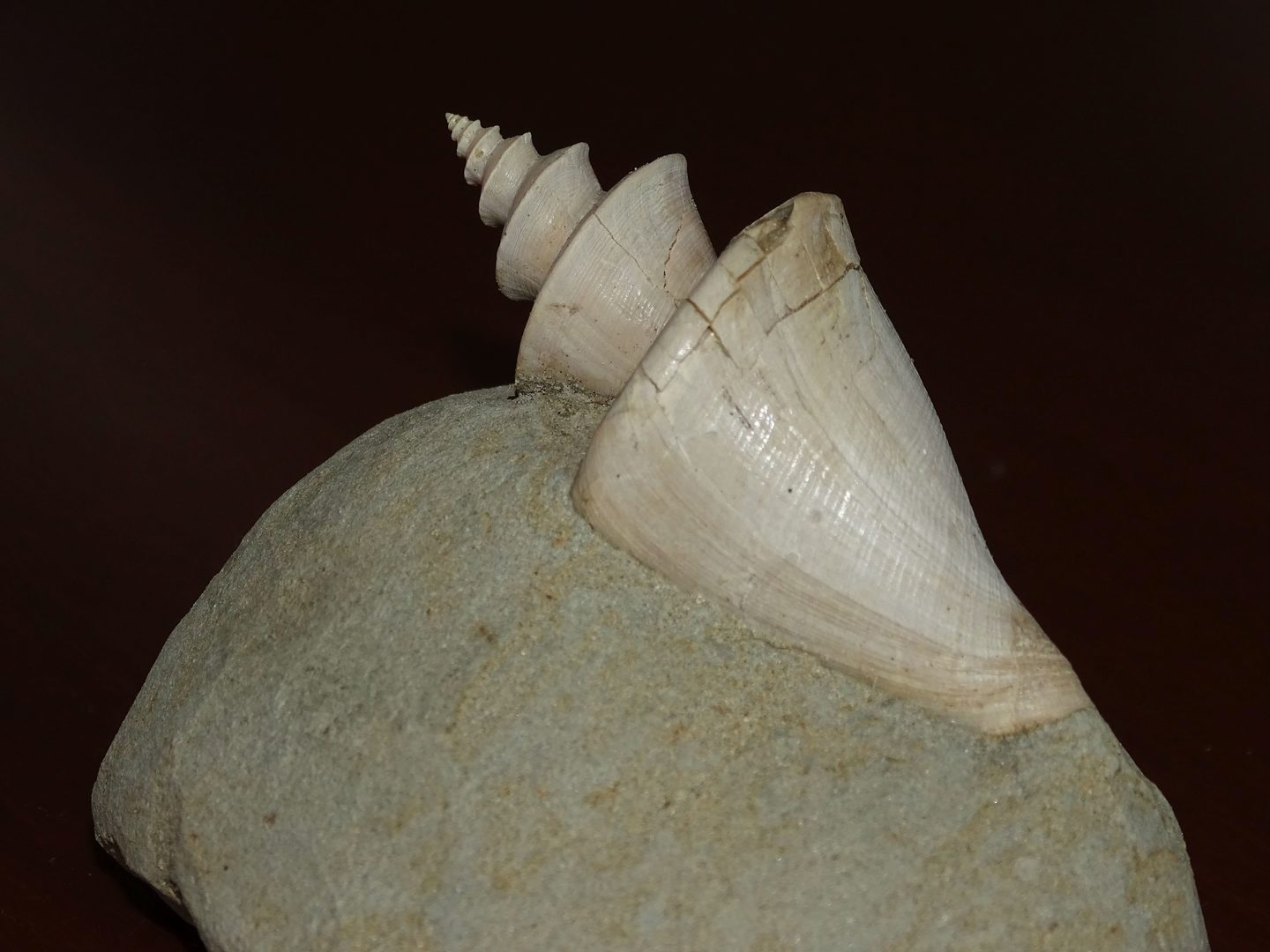
6.2 Onna-son/OIST Children’s School of Science 2017
- Date: August 21-25, 2017
- Venue: Fureai Taiken Center
- Speaker (among others): Bernd Kuhn
- Topic: Palaeontology of Okinawa for Grade 1-3
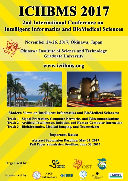
6.3 ICIIBMS 2017
- The Second International Conference on Intelligent Informatics and BioMedical Sciences (ICIIBMS) is in the planning phase.
- Date: November 24-26, 2017
- Venue: OIST
- Organized by researchers of the University of the Ryukyus, the Okinawa National College of Technology, and OIST
- 140 participants from 15 countries around the world
6.4 Prof. C. Justin Lee (KIST, South Korea)
- Title:Targeting GABA from reactive astrocytes to treat Alzheimer's disease
- Date: September 22, 2017
6.5 Dr. Masanori Murayama (RIKEN BSI, Japan)
- Title: Top-Down Cortical Circuit for Perception and Memory Consolidation in Mice
- Date: December 12, 2017
7. Other
7.1 Planning of ICIIBMS 2018
- The Third International Conference on Intelligent Informatics and BioMedical Sciences (ICIIBMS) is in the planning phase.
- Date: October 21-24, 2018
- Venue: Bangkok
- Organized by researchers of the University of the Ryukyus, the Okinawa National College of Technology, KMITL (Bangkok, Thailand), and OIST