FY2015 Annual Report
Optical Neuroimaging Unit
Associate Professor Bernd Kuhn
Abstract
In the FY 2015 the Optical Neuroimaging Unit continued several imaging projects. Most research projects focused on imaging neuronal activity in the mouse brain.
1. Staff
- Dr. Bernd Kuhn, Associate Professor
- Dr. Akihiro Funamizu, Postdoc (shared with Doya Unit)
- Dr. Christopher J. Roome, Postdoc
- Dr. Sigita Augustinaite, Postdoc
- Dr. Shinobu Nomura, Postdoc
- Ray X. Lee, Ph.D. student
- Neil Dalphin, Ph.D. student
- Leonidas Georgiou, Ph.D. student
- Dr. Kazuo Mori, Technical Assistant
- Hiroko Chinone, Research Unit Administrator
2. Collaborations
2.1 Neural substrate of dynamic Bayesian inference in the cerebral cortex
- Dynamic Bayesian inference allows a system to infer the environmental state under conditions of limited sensory observation. We use in vivo two-photon calcium imaging and probabilistic population decoding to show that cortical neurons in layers 2, 3, and 5 implement the fundamental features (prediction and updating) of Dynamic Bayesian inference.
- Type of collaboration: Joint research
- Researchers:
- Professor Dr. Kenji Doya, Neural Computation Unit, OIST
- Dr. Akihiro Funamizu, Neural Computation Unit and Optical Neuroimaging Unit, OIST
3. Activities and Findings
3.1 Neural substrate of dynamic Bayesian inference in the cerebral cortex
Our brain often receives limited sensory information to understand the state of the outside world. Therefore the ability to estimate the current state through mental simulation is essential. In a stochastic dynamic environment, this state estimation is realized by dynamic Bayesian inference, such as Kalman filtering. To investigate the neural substrate of dynamic Bayesian inference, we use two-photon imaging in awake behaving mice which enables us to simultaneously image multiple identified neurons and their calcium activities while the mouse conducts an auditory virtual navigation task.
A mouse is head restrained and maneuvers a spherical treadmill. 12 speakers around the treadmill provide an auditory virtual environment. The direction and amplitude of sound pulses emulate the location of the sound source, which is moved according to the mouse’s locomotion on the treadmill. When the mouse reaches the sound source (goal) and licks a spout, it gets a water reward. The task consists of two conditions: continuous condition in which the guiding sound is presented continuously and intermittent condition in which the sound is presented intermittently.
In the task, mice increase licking as they approached the goal both with and without cue sounds. The anticipatory licking is disturbed selectively during sound suspension by inactivating posterior parietal cortex (PPC) with the GABA-A receptor agonist muscimol. These results suggest that mice estimate the goal distance based on their own actions and that PPC is involved in the estimation.
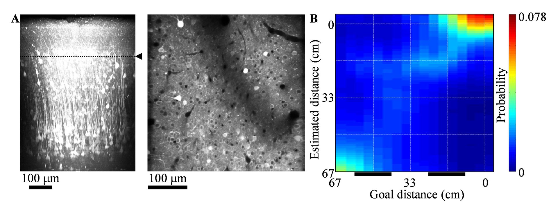
Calcium is an important second messenger in neurons and an increased calcium concentration is correlated with neuronal activity. For this reason we use the genetically encoded calcium indicator GCaMP6f in PPC and adjacent posteromedial cortex (PM) of mice to detect neuronal activities with two-photon microscopy. We record the activities of 200 – 500 neurons simultaneously in each of layers 2, 3 and 5 for one hour continuously (Figure). By decoding the population activity using machine learning techniques, we find that some neurons represented goal distances during sound suspension (prediction), more robustly in PPC than in PM and in layers 3, 5 more robustly than in layer 2. Uncertainty of prediction decreased with observation of cue sounds (updating). These results suggest that cortical microcircuits implement dynamic Bayesian inference for state estimation and that PPC takes a role in the estimation of target location using an action-dependent state transition model.
3.2 Voltage imaging with two-photon microscopy
Many neurons have elaborate dendrites with a wide variety of ion channels governing their electric properties. Unfortunately, it is very difficult to record the voltage in dendrites due to their fine structure. At the same time dendritic voltage is a key to understand neuronal information processing and therefore also to understand brain function.
We develop tools to measure dendritic voltage changes in vivo. We use the pure electrochromic voltage sensitive dyes ANNINE-6 (Kuhn & Fromherz 2003) and ANNINE-6plus (Fromherz et al 2008). Pure electrochromic means that the change of fluorescence only depends on the electric field over the dye molecule and that there are no other, slower effects modulating the spectra.
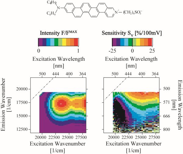
Figure: ANNINE-6 (top) is a pure electrochromic or pure Stark-shift probe. This can be shown by analyzing the spectral shift in excitation and emission caused by a change of the electric field over the dyes (Kuhn & Fromherz 2003).
We label single neurons in vivo through an access port in the chronic cranial window (Roome & Kuhn 2014).
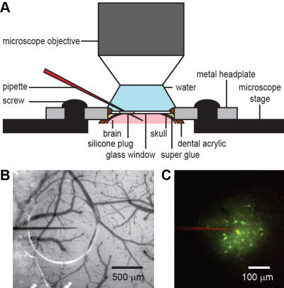
Figure: A. A chronic chranial window with access port allows to inject drugs or dyes repeatedly into the brain. It also allows electrical recordings. B. A beveled pipette can penetrate the membrane of the access port and C it can be used to inject dye locally into the brain (Roome & Kuhn 2014)
By excitation at the red edge of the a absorption spectrum we can reduce phototoxicity and increase voltage sensitivity dramatically (Kuhn et al 2004).
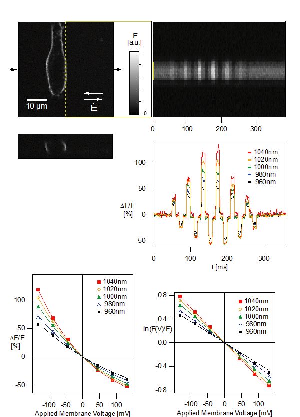
Figure: HEK293 cells labeled with ANNINE-6 are imaged with two-photon microscopy (top left). A line-scan is performed while alternating extracellular electric fields over the cell are applied (top right). The relative fluorescence change per applied voltage increases toward the spectral edge of the absorption spectrum of ANNINE-6 (bottom). (Kuhn et al 2004)
After filling a cell we can electrically record from the soma and image dendritic voltage changes in vivo.
3.3 Imaging thalamo-cortical interaction
Sensory information is relayed through the thalamus to cortex. At the same time, cortex is regulating thalamic activity by driving and modulatory feedback. We try to image these interactions.
3.4 Imaging PKA activity in vivo
Protein kinase A (PKA) is an important encym in the signaling cascade connecting neuronal activity and expression and modulation of proteins. We use genetically encoded indicators to image PKA activity in vivo.
3.5 pH measurement within protein crystals
Protein crystals can be considered nano porous materials. We measure the conditions inside the nano pores with the help of molecular probes. We use pH-sensitive dyes, load them into the crystals, and record pH changes in response to pH changes of the bath. We find differences between the pH of the bath and the nano pores. We try to find a model that explains this difference.
4. Publications
4.1 Journals
Nothing to report
4.2 Books and other one-time publications
Nothing to report
4.3 Oral and Poster Presentations
- A. Funamizu, B. Kuhn, K. Doya. Action-dependent state prediction in mouse posterior parietal cortex during an auditory virtual navigation task, ICIIBMS 2015, Onna-son, Japan (28-30 Nov. 2015).
- C.J. Roome and B. Kuhn. A method for exploring input-output relations of neurons in awake mice, ICIIBMS 2015, Onna-son, Japan (28-30 Nov. 2015).
- R.X. Lee, G.J. Stephens, B. Kuhn. Prediction of spontaneous behavioral dynamics by neuronal population imaging in mouse neocortex, ICIIBMS 2015, Onna-son, Japan (28-30 Nov. 2015).
- A. Funamizu, B. Kuhn, K. Doya. Action-dependent state prediction in mouse posterior parietal cortex during an auditory virtual navigation task, SfN, Chicago, U.S.A. (28-30 Nov. 2015).
- O. Jaidar, C.J. Room, Y. Nakano, M. Garcia-Munoz, B. Kuhn, G. Arbuthnott. Thalamocortical axons and deep cortical layer dendritic arbor in motor cortex layer I display a dynamic array of calcium transients during locomotion and systemic blockade of dopamine receptors, BNA 2015 Festival of Neuroscience, Edinburgh, G.B. (12-15. April 2015).
- C.J. Roome and B. Kuhn. A method for exploring input-output relations in awake mice. New Advances in Optical Imaging of Live Cells and Organs, Cold Spring Harbor Asia Conference. Organizer: Prof. G. Bi., W. Gan, A. Konnerth, A. Kusumi. Suzhou, China (7-11. Dec. 2015).
- B. Kuhn. Imaging neuronal activity in awake mice. Japanese Society of Neurogastroenterology. Organizer: Prof. Koji Yakabi. OIST, Japan (12-13. Nov. 2015).
- B. Kuhn. Voltage imaging with two-photon microscopy. Invited by Prof. Lawrence B. Cohen. Center for Functional Connectomics, KIST, South Korea (4. Nov. 2015).
- B. Kuhn. Two-photon microscopy - A tool for many different applications. Invited by Prof. C.-T. Yen. Department of Life Science, National Taiwan University, Taipei, Taiwan (8. Sep. 2015).
- B. Kuhn. Voltage imaging with two-photon microscopy. RIKEN BSI Neuroscience Symposium 2015. Organizers: Prof. D. Van Vactor, Prof. Jeffrey Wickens. RIKEN BSI, Wako-shi, Saitama, Japan (31. July- 1. August 2015).
5. Intellectual Property Rights and Other Specific Achievements
Nothing to report
6. Meetings and Events
6.1 Workshop: Okinawa Computational Neuroscience Course 2015
- Date: June 16- July 3, 2014
- Venue: OIST Seaside House
- Organizers: Drs. E. De Schutter, K. Doya, J. Wickens
- Speaker (among others): Bernd Kuhn
- Title 1: Voltage-gated channels and the Hodgkin-Huxley model of neuronal activity
- Title 2: Functional Optical Imaging Methods
6.2 Onna-son/OIST Children’s School of Science 2015
- Date: August 18-22, 2014
- Venue: Fureai Taiken Center
- Speaker (among others): Bernd Kuhn
- Topic: Palaeontology of Okinawa for Grade 1-3
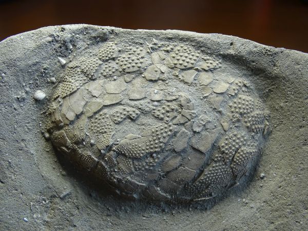
Figure: Fossil sea urchin of Okinawa found in late Tertiary marine sediments (about 2 M years old).
6.3 International Undergraduate Collaborative Workshop
- Date: 10-14 August 2015
- Organizer: OIST Graduate School
- Venue: OIST, Onna-son
- Instructors (among others): Bernd Kuhn
- Topic: Unpacking animal behavior, neural circuit function and neuronal cell biology through interdisciplinary and quantitative analysis
6.4 7th Research Area Meeting, Grant in Aid for Scientific Research on Innovative Areas
- Date: 25. April 2015
- Organizer: Prof. K. Doya
- Tutorial speaker (among others): Bernd Kuhn
- Topic: Elucidation of the Neural Computation for Prediction and Decision Making
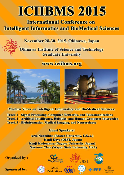
6.5 ICIIBMS 2015
- First International Conference on Intelligent Informatics and BioMedical Sciences (ICIIBMS) was held at the OIST Conference Center. Over 150 people from 18 countries participated.
It was also very nice that researchers not only from academia but also from industry attended. - Date: November 28-30, 2015
- Venue: OIST
- Organized by researchers of the University of the Ryukyus, the Okinawa National College of Technology, and OIST
7. Other
7.1 Hosting the German Consul General and a German Delegation with the Chairman of the CDU/CSU
- Hosting the visit of Mr. Volker KAUDER, Chairman of the CDU/CSU, Mrs. Marie-Luise DÖTT, Environmental Policy Spokeswoman for CDU/CSU, Dr. Andreas MOM, Chief of Staff to the Chairman/Personal Assistant to the Chairman, CDU/CSU, Mr. Ulrich SCHARLACK, Press Spokesman for CDU/CSU, Dr. Michael Frank, Foreign Policy and Security Adviser to the Chairman, CDU/CSU, Paul LINNARZ, Regional Representative for Economic Policy / President Representative for Japan, Konrad-Adenaurer-Stiftung, Dr. Ingo Karsten, Consul General in Osaka-Kobe, and Ms. Tamaki Asukai
- Date: 21. March 2016
7.2 Planning of ICIIBMS 2017
- The Second International Conference on Intelligent Informatics and BioMedical Sciences (ICIIBMS) is in the planning phase.
- Date: November 24-26, 2017
- Venue: OIST
- Organized by researchers of the University of the Ryukyus, the Okinawa National College of Technology, and OIST