

FY2013 Annual Report
Developmental Neurobiology Unit
Associate Professor Ichiro Masai

Abstract
The vertebrate neural retina is derived from the ventral region of the forebrain. In this region, six classes of neurons differentiate and form the neural circuit underlying visual transduction. Thus, the retina provides an excellent model for studying cell differentiation and neural circuit formation in the vertebrate brain. Furthermore, more than one hundred hereditary retinal diseases causing photoreceptor degeneration have been identified in humans. Understanding the pathological processes of photoreceptor degeneration is an important issue from a medical perspective. We are currently investigating the mechanisms underlying retinal cell differentiation and photoreceptor degeneration, using zebrafish as an animal model. Regarding the project on retinal cell differentiation, in 2013, we showed that retinal neurogenesis is delayed in zebrafish stem-loop binding protein 1 (slbp1) mutant. Because SLBP1 regulates a replication-dependent synthesis and degradation of histone protein, this finding suggests that replication-coupled histone regulation is important for retinal neurogenesis. Furthermore, to examine cell-cycle regulation with high accuracy, we visualized cell-cycle phases using zebrafish fluorescent ubiquitination-based cell cycle indicator (Fucci). We applied this Fucci technique to zebrafish lens epithelium and revealed a spatial regulation of cell proliferation in zebrafish lens epithelium. Regarding the project on photoreceptor degeneration, in 2013, we examined zebrafish b-SNAP mutant showing photoreceptor degeneration. We showed that a BH3-only SNARE, BNip1, mediates photoreceptor apoptosis in absence of b-SNAP activity. We propose that BNip1 functions as an alarm that senses vesicular fusion defects and induces apoptosis.
1. Staff
- Dr. Yuko Nishiwaki, Group leader
- Dr. Toshiaki Mochizuki, Researcher
- Dr. Junichi Kawada, Researcher (– March 2014)
- Dr. Maria Iribarne, Researcher
- Ms. Asuka Yoshizawa, Technician (– June 2013)
- Mr. Yutaka Kojima, Technician
- Mr. Shohei Nakamura, Technician (– October 2013)
- Ms. Eri Oguri, Technician
- Ms. Ayako Matsuzaki, Technician (May 2013 – March 2014)
- Mr. Masato Araragi, Technician (September 2013 –)
- Ms. Miyuki Suenaga, Technician (April 2014 –)
- Ms. Yayoi Tomoyose, Research Assistant
- Ms. Kazumi Toguchi, Research Assistant
- Ms. Yuko Yamazato, Research Assistant (– December 2013)
- Ms. Moe Inafuku, Research Assistant (December 2013 –)
- Ms. Chitose Mizuta, Research Assistant (June 2014 –)
- Dr. Mariko Kinoshita-Kawada, Research Assistant (– March 2014)
- Ms Ayako Gima, Research Administrator/Secretary
2. Collaborations
- Theme: In vivo functional analysis of hypoxia response genes using the zebrafish retina
- Type of collaboration: Joint research
- Researchers:
- Prof. Ichiro Masai (Developmental neurobiology unit, OIST)
-
Prof. Masayuki Matsushita (Department of Medicine, Ryukyu University)
3. Activities and Findings
3.1 Mechanism regulating retinal neurogenesis in zebrafish
The vertebrate retina is initially specified in the anterior neural plate and evaginates from the ventral region of the forebrain as the optic cup. In this region, six major classes of retinal neurons and one class of glial cells differentiate to form the neural circuit that mediates phototransduction and visual processing (Fig. 1A). Thus, the retina provides an excellent model for studying the mechanisms underlying cell differentiation and neural circuit formation in the developing brain.
In the developing retina, retinal cells initially proliferate. At a particular developmental stage, retinal cells start to exit from cell cycle and differentiate into neurons, whose process is called neurogenesis. We have examined molecular mechanism that regulates retinal neurogenesis in zebrafish. Previously, we identified several signaling molecules, Hedgehog, Fgf, Wnt, Notch, Histone deacetylase 1, and a cell-polarity regulator MAGUK p55 superfamily number 5 (MPP5, zebrafish nagie oko), which cooperatively regulate the balance between proliferation and differentiation during retinal neurogenesis (Fig. 1B). However, molecular mechanisms that coordinate cell proliferation and neurogenesis are not fully understood.
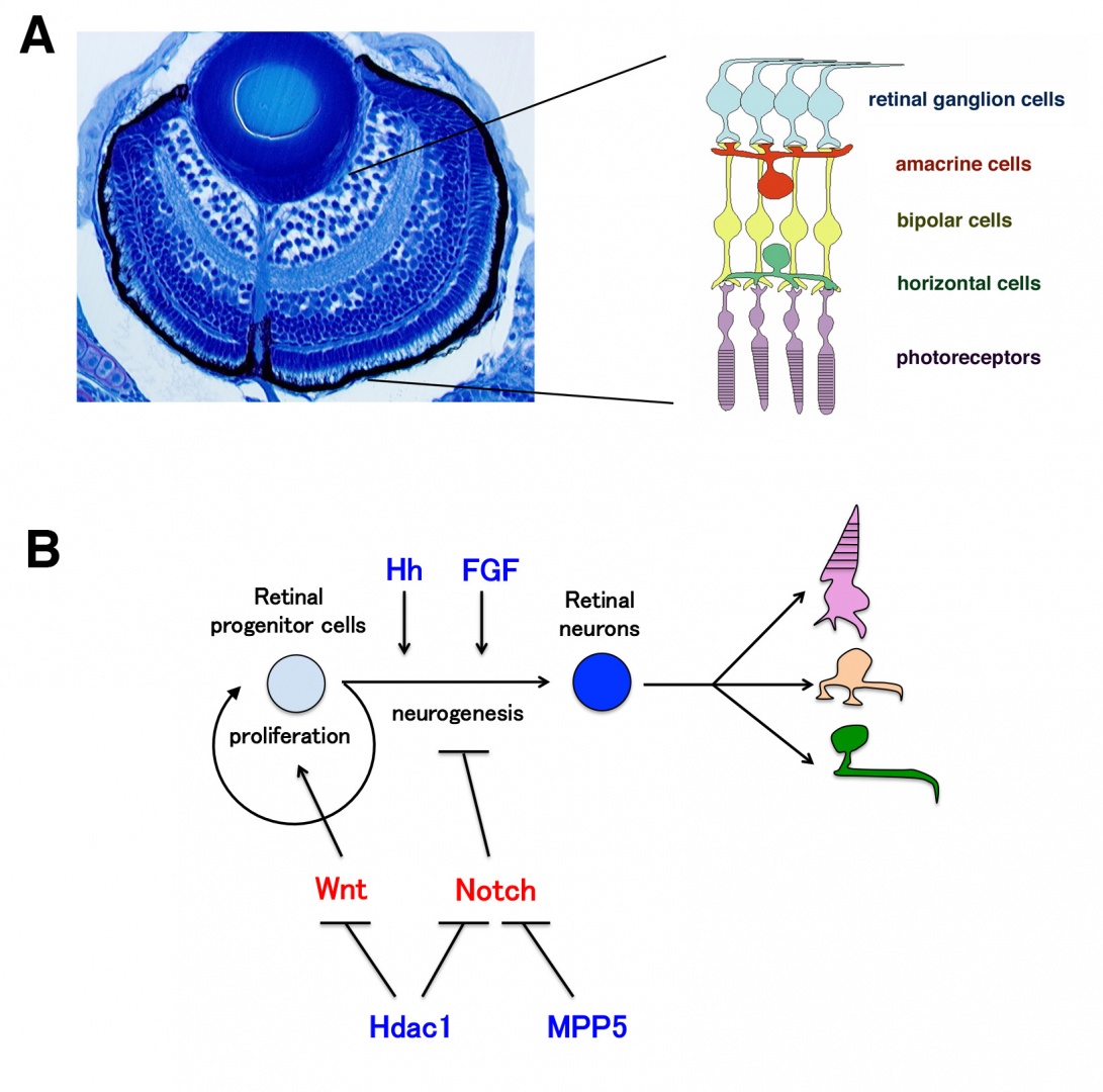
Figure 1: Zebrafish retina and signaling network regulating retinal neurogenesis. (A) Plastic section of 3 dpf zebrafish retina. Six major classes of retinal neuron including photoreceptors differentiate and form layered structures. (B) FGF and Hh regulate the induction and progression of retinal neurogenesis in zebrafish. Wnt and Notch signaling pathways promote cell differentiation and neurogenic inhibition, respectively. Hdac1 promotes retinal neurogenesis by suppressing both Wnt and Notch signaling pathways. A cell polarity regulator MPP5 (nagie oko) is required for proper timing of neurogenesis through suppression of Notch signaling.
In 2013, we found that retinal neurogenesis is delayed in zebrafish stem-loop binding protein 1 (slbp1) mutant. SLBP1 binds to a stem-loop structure at the 3’-end of histone mRNAs, and regulates a replication-dependent synthesis and degradation of histone protein. We found that retinal cell proliferation becomes slow in the slbp1 mutant, resulting in cessation of retinal stem cell proliferation. Although retinal stem cells stop proliferation at 2 days post-fertilization (dpf), retinal progenitor cells in the central retina continue to proliferate and generate retinal neurons until 5 dpf (Fig. 2A). We found that this progenitor proliferation depends on Notch signaling, suggesting that Notch signaling maintains retinal progenitor proliferation when faced with reduced SLBP1 activity. Thus, SLBP1 cooperates with Notch singling to promote cell proliferation and subsequent neurogenesis in zebrafish retina (Fig. 2B). These data suggest that SLBP1 constitutes one of core components that regulate retinal neurogenesis.
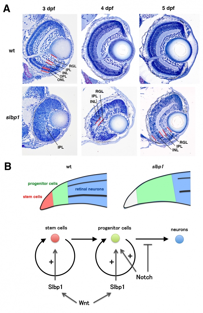
Figure 2: Retinal neurogenesis is delayed in zebrafish slbp1 mutant (A) Sections of wild-type and the slbp1 mutant retinas at 3 – 5 dpf. In wild-type retina, all layers are formed after 3 dpf. However, in the slbp1 mutant retina, retinal lamination is not observed, except a small IPL at 3 dpf. In the slbp1 mutant retina, the IPL becomes large, and the RGL and INL are distinct at 4 dpf. In the slbp1 mutant retina, the RGL, IPL, and INL are more evident, but the ONL is absent at 5 dpf. RGL, retinal ganglion cell layer; IPL, inner plexiform layer; INL, inner nuclear layer; OPL, outer plexiform layer; ONL, outer nuclear layer. (B) (Upper) Schematic drawing of phenotypes for retinal cell proliferation and neurogenesis in wild type, and slbp1 mutant. In wild-type retina, areas of retinal stem cells (red), retinal progenitor cells (green), and differentiated neurons (blue) are specified along the peripheral-central axis. In the slbp1 mutant, retinal stem cells stop cell proliferation (white), retinal progenitor cell proliferation is slow, and retinal neurogenesis is delayed. (Bottom) SLBP1 is required for retinal stem cell proliferation. SLBP1 cooperates with Notch signaling to promote retinal progenitor cell proliferation.
3.2 Visualization of cell-cycle phases with fluorescent proteins using Fucci
The first step of neurogenesis is the exit from the cell cycle. Previously we showed that the earliest neurogenic marker, ath5 (also designated as atoh7), is initiated in the G2 phase in retinal progenitor cells just prior to the final cell division, which generates two neuronal daughter cells. Thus, it is likely that the commitment to the retinal neuron is associated with cell-cycle progression. To accurately understand the relationship between cell proliferation and cell differentiation, we visualized cell-cycle phases differentially with fluorescent proteins, using the zebrafish Fucci (fluorescent ubiquitination-based cell cycle indicator) (Sugiyama et al., 2009, Proc Natl Acad Sci USA 106, 20812–20817). In the Fucci system, the cell-cycle dependent expression of two cell-cycle regulators, Geminin and Cdt1, are visualized with fluorescent proteins fused to their regulatory peptide sequences. We generated the zebrafish transgenic line carrying genes encoding mCherry-tagged Geminin and green fluorescent protein (GFP)-tagged Histone 2A. In this transgenic fish, we were able to clearly distinguish three different phases in the developing retina as well as the lens; the G1, S-G2, and M phases (Fig. 3A). Furthermore, we were able to observe chromosome condensation in the M phase (Fig. 3B) and measure the orientation of cell division from in vivo time-lapse imaging data.
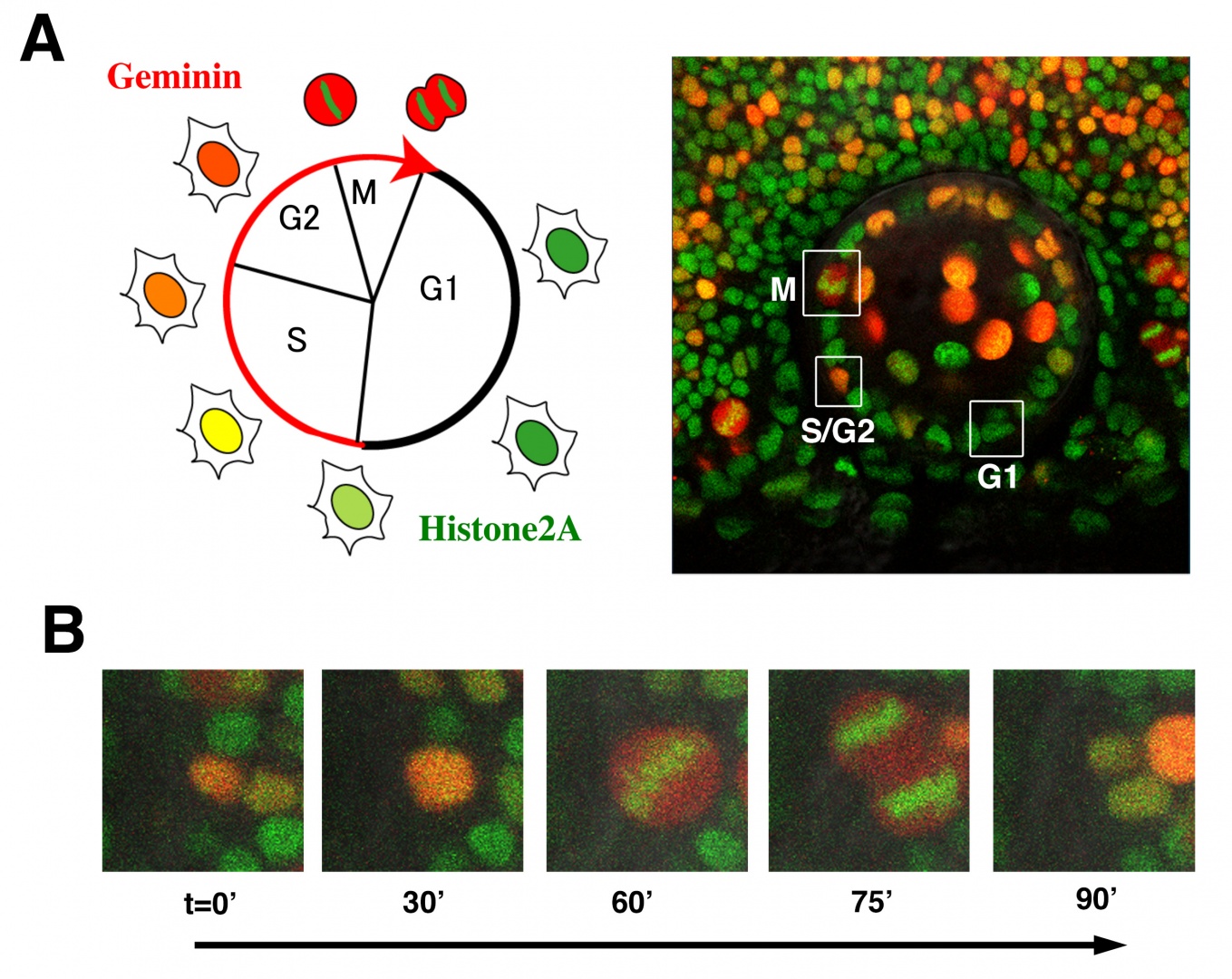
Figure 3: Visualization of cell-cycle progression in the zebrafish retina and lens. (A) (Left panel) Relationship between cell cycle phases and fluorescent protein expression. Geminin (red) are expressed exclusively in S/G2/M phase. GFP-tagged Histone2A (green) labels nuclei. (Right image) Confocal images of zebrafish transgenic line carrying GFP-tagged Histone 2A (green) and mCherry-tagged Geminin (red). Cells in the G1, S/G2, and M phase are distinguished. (B) Confocal images of mitosis in zebrafish retinal cells line with GFP-tagged Histone 2A (green) and mCherry-tagged Geminin (red) transgenes. GFP-tagged Histone2A expression (green) becomes condensed in segregating chromosomes.
In 2013, using this transgenic fish, we observed the cell-cycle progression, cell division, and cell migration in the zebrafish lens epithelium using time-lapse techniques, because the lens epithelium is more easily accessed than the neural retina (Fig. 4A). We found that cell proliferation was highly active in a marginal zone just anterior to the equator in the lens epithelium. Furthermore, cell-division orientation was biased longitudinally in the anterior region of the lens epithelium, whereas it was circumferentially biased in the peripheral proliferating zone (Fig. 4B). These data suggest that cell proliferation and cell division are spatially patterned in the zebrafish lens epithelium (Fig. 4C). In many developing tissues, it has been reported that cells tend to divide along their long axis. This is referred to as “the Hertwig rule”. We found that the long axis was similarly biased longitudinally and circumferentially in the anterior and proliferating region in the zebrafish lens epithelium, respectively (Fig. 4D), suggesting that lens epithelial cells follow the Hertwig rule. These observations suggest that apical cell geometry correlates with cell division orientation in the lens epithelium.
Since apical cell geometry depends on cell adhesion and a cell adhesion molecule E-cadherin is expressed in the lens epithelium, we examined cell-division orientation and apical cell geometry in lens epithelium of zebrafish e-cadherin mutant. Cell-division orientation and cell geometry were more longitudinally biased in lens epithelium of e-cadherin mutant than in wild type (Fig. 4E). These data suggest that E-cadherin regulates the spatial pattern of cell division orientation through epithelial cell geometry in lens epithelium.
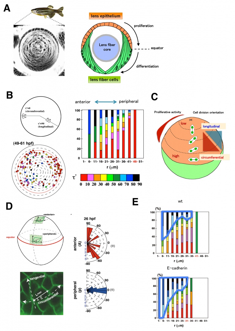
Figure 4: Cell proliferation and division are spatially regulated in the zebrafish lens. (A) (Left) Zebrafish lens labeled with fluorescence lipid dye. (Right) Anterior lens epithelial cells (orange) are proliferative and start to differentiate into lens fiber cells (green) at the equator. Differentiating lens fiber cells elongate to cover the old lens fiber core (blue). (B) (Left) Plotting of mitosis position on anterior projection view of lens epithelium. Cell division was more frequent in the marginal zone anterior to the equator. Angles of cell-division orientation relative to the circumferential axis are indicated with color tone spectrum from red (0 degree) to blue (90 degree). (Right) Histogram of cell division orientation. Cell division orientation was biased longitudinally ands circumferentially in the anterior and peripheral proliferating zone, respectively. (C) Schematic drawing of pattern of cell proliferation and cell division orientation in the zebrafish lens epithelium. (D) (Left bottom) Labeling of lens epithelium at the proliferating zone with a fluorescent lipid dye (green), which visualizes cell membranes. (Right) Histogram of angle of cell division to the circumferential axis. A majority of cell divisions are in parallel with the longitudinal and circumferential axis in the anterior and peripheral region, respectively. (E) Histogram of cell division orientation of wild-type and e-cadherin mutant lens epithelium. The fraction of longitudinally oriented cell division increases in e-cadherin mutant.
3.3 Mechanisms underlying photoreceptor degeneration
Photoreceptors are highly specialized neurons that efficiently detect light stimuli. To date, more than a hundred genes associated with inherited photoreceptor degeneration have been identified in humans (see the homepage of the Retinal Information Network at http://www.sph.uth.tmc.edu/Retnet/). However, the mechanisms underlying the pathological processes involved in photoreceptor degeneration remain to be elucidated. To determine the mechanisms underlying photoreceptor degeneration, we have screened zebrafish mutants with dysfunctional visual behavior by testing the optokinetic response. We identified several zebrafish mutants, in which photoreceptors differentiate but later degenerate. In 2010, we reported that one of these mutants carried a genetic mutation in cGMP-phosphodiesterase 6c (PDE6c), a mediator molecule of cone-specific phototransduction, suggesting a link between achromatopsia and progressive cone dystrophy. In 2012–2013, we examined another mutant, coa, whose photoreceptors rapidly undergo apoptosis before maturation (Fig. 5A). We found that the coa mutant gene encodes b-soluble N-ethylmaleimide-sensitive factor attachment protein (b-SNAP) (Fig. 5B). b-SNAP regulates the fusion of transport vesicles to intracellular membrane organelles. These observations suggest that vesicular transport defects cause photoreceptor apoptosis in zebrafish.
Mitochondria-dependent apoptosis is promoted by pro-apoptotic Bcl2 family proteins, Bax, and inhibited by anti-apoptotic Bcl2 family proteins, Bcl2. BH3-only proteins promote apoptosis by modulating the balance between pro-apoptotic and anti-apoptotic Bcl2 proteins. We found that a BH3-only protein, BNip1, mediates the coa-mediated photoreceptor apoptosis (Fig. 5C). BNip1 is a component of the syntaxin 18 SNARE complex, and the syntaxin 18 SNARE complex regulates retrograde transport from the Golgi apparatus to the ER. b-SNAP promotes the recycle of vesicular fusion machinery, SNARE, by disassembling the cis-SNARE complex generated by vesicular fusion. Thus, it is likely that the syntaxin 18 cis-SNARE complex is accumulated in the coa mutant. We found that failed disassembly of the syntaxin 18 cis-SNARE complex activates the BNip1-dependent photoreceptor apoptosis in the coa mutant. These data suggest that the syntaxin 18 cis-SNARE complex monitors vesicular fusion competence and that BNip1 transforms vesicular fusion defects into apoptosis (Fig. 5D). From these data, we propose that BNip1 functions as an alarm that monitors abnormalities of vesicular fusion and transforms vesicular fusion defects into apoptosis in photoreceptors.
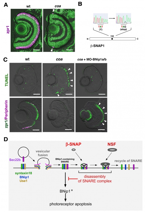
Figure 5: BNip1 mediates photoreceptor apoptosis in response to vesicular fusion defects. (A) Zpr1 antibody labeling of 6 dpf wild-type and coa mutant retinas. All nuclei are counterstained with Sytox-Green (green). Arrowheads indicate zpr1 expression (magenta) in the CMZ of the coa mutant. (B) A non-sense mutation occurs at 152Y in b-SNAP gene in the coa mutant. (C) (upper panels) TUNEL (green) of 84 hpf wild-type and coa mutant retinas, and coa mutant retinas injected with a mixture of BNip1a and BNip1b morpholino anti-sense oligos. Arrowheads indicate apoptosis in the ONL. (lower panels) Zpr1 labeling (green) and GFP-tagged Peripherin (magenta) in 84 hpf wild-type, and coa mutant retinas, and coa mutant retinas injected with a mixture of BNip1a and BNip1b morpholino anti-sense oligos. Arrowheads indicate rescued photoreceptors. (D) BNip1 is a component of the syntaxin18 SNARE complex, which regulates retrograde transport from Golgi to ER. Vesicular fusion generates the cis-SNARE complex, which is subsequently disassembled by SNAP and NSF for SNARE recycle. In the coa mutant, -SNAP activity is reduced, resulting in the accumulation of the syntaxin 18 cis-SNARE complex, which activates BNip1 pro-apoptotic activity. We propose that the syntaxin18 cis-SNARe complex functions as an alarm that monitors vesicular fusion competence and that BNip1 transforms vesicular fusion defects into photoreceptor apoptosis. Scale bars: 50 micro-m.
4. Publications
4.1 Journals
Imai, F., Yoshizawa, A., Matsuzaki, A., Oguri, E., Araragi, M., Nishiwaki, Y. & Masai, I. Stem-loop binding protein is required for retinal cell proliferation, neurogenesis, and intraretinal axon pathfinding in zebrafish. Developmental biology, doi:10.1016/j.ydbio.2014.07.020 (2014).
Mochizuki, T. & Masai, I. The lens equator: a platform for molecular machinery that regulates the switch from cell proliferation to differentiation in the vertebrate lens. Development, growth & differentiation 56, 387-401, doi:10.1111/dgd.12128 (2014).
Nishiwaki, Y., Yoshizawa, A., Kojima, Y., Oguri, E., Nakamura, S., Suzuki, S., Yuasa-Kawada, J., Kinoshita-Kawada, M., Mochizuki, T. & Masai, I. The BH3-only SNARE BNip1 mediates photoreceptor apoptosis in response to vesicular fusion defects. Developmental cell 25, 374-387, doi:10.1016/j.devcel.2013.04.015 (2013).
4.2 Books and Other One-Time Publications
Nishiwaki, Y., Masai, I. The BH3-only SNARE BNip1 mediates photoreceptor apoptosis in response to vesicular fusion defects. First Author’s, http://first.lifesciencedb.jp, © 2013 (2013).
4.3 Oral and Poster Presentations
(Oral, International conference)
Nishiwaki, Y., and Masai, I. Molecular mechanism that links vesicular fusion defects and photoreceptor degeneration, in 6th Asia Oceania Zebrafish Meeting, Hong Kong, China (2014).
Mochizuki, T., Luo, Y.-J., Suzuki, S., and Masai, I. . E-cadherin-dependent cell adhesion regulates cell migration pattern in the zebrafish lens epithelium, in 11th International Conference on Zebrafish Development and Genetics, Madison, USA (2014).
Nishiwaki, Y., Yoshizawa, A., Kojima, Y., Oguri, E., Nakamura, S., Suzuki, S., Yuasa-Kawada, J., Kinoshita-Kawada, M., Mochizuki, T. and Masai, I. Syntaxin18 cis-SNARE complex is a novel alarm factor that detects vesicular transport defects, in 8th European Zebrafish Meeting, Barcelona, Spain (2013).
Mochizuki, T., Suzuki, S., Sakaue-Sawano, A., Miyawaki, A. and Masai, I. Cadherins regulate cell geometry and cell division orientation in zebrafish lens epithelium, in 8th European Zebrafish Meeting, Barcelona, Spain (2013).
(Poster, international conferences)
Nishiwaki, Y., Nakamura, S., Oguri, E., Araragi, M., Kojima, Y., and Masai, I. . Molecular mechanism that converts vesicular fusion defects into apoptosis in photoreceptors, in 11th International Conference on Zebrafish Development and Genetics, Madison, USA (2014).
Nishiwaki, Y., Nakamura, S., Oguri, E., Araragi, M., Kojima, Y. and Masai, I. . Molecular Mechanism that converts vesicular fusion defects into apoptosis in photoreceptors, in Gordon research seminar – Visual System Development, Lucca, Italy (2014).
Nishiwaki, Y., Nakamura, S., Oguri, E., Araragi, M., Kojima, Y. and Masai, I. . Molecular mechanism that converts vesicular fusion defects into apoptosis in photoreceptors, in Gordon Research Conference –Visual System Development, Lucca, Italy (2014).
Nishiwaki, Y., Nakamura, S., Oguri, E., Kojima, Y., Suzuki, S., Prescott, J., and Masai, I. Molecular mechanism underlying photoreceptor cell death in response to genetic mutations of rhodopsin gene, in 6th Asia Oceania Zebrafish Meeting, Hong Kong, China (2014).
(Invited talks)
Masai, I. Visualization of cell proliferation and migration in zebrafish lens epithelium, in Gordon Research Conference –Visual System Development, Lucca, Italy (2014).
Nishiwaki, Y. a. M., I. Molecular mechanism underlying photoreceptor degeneration in response to vesicular fusion defects, in 20th East Asia Joint Symposium on “Global Medical Science for the 22nd Century, Tokyo, Japan (2013).
Mochizuki, T. a. M., I. Spatial regulation of cell proliferation and differentiation in the zebrafish lens, in The 46th Annual Meeting of the Japanese Society of Developmental Biologists, Matsue, Shimane, Japan (2013).
Masai, I. Molecular mechanism underlying photoreceptor degeneration in response to vesicular fusion defects, in Imaging Symposium, Nikon Imaging Center, Harvard Medical School, Boston, USA (2013).
Masai, I. Molecular mechanism underlying photoreceptor degeneration in response to vesicular fusion defects, in Asia ARVO 2013, New Delhi, India (2013).
(Oral, domestic conferences)
Mochizuki, T., Luo, Y.-J., Suzuki, S., and Masai, I. E-cadherin-dependent cell adhesion regulates cell migration pattern in the zebrafish lens epithelium, in The 47th Annual Meeting of the Japanese Society of Developmental Biologists, Nagoya, Japan (2014).
Nishiwaki, Y., Yoshizawa, A., Kojima, Y., Oguri, E., Nakamura, S., Suzuki, S., Yuasa-Kawada, J., Kinoshita-Kawada, M., Mochizuki, T. and Masai, I. Syntaxin18 cis-SNARE complex is a novel alarm factor that detects vesicular transport defects, in 46th Annual Meeting of the Japanese Society of Developmental Biologists, Matsue, Shimane, Japan (2013).
Mochizuki, T., Suzuki, S., Sakaue-Sawano, A., Miyawaki, A., and Masai, I. . Cadherin-mediated physical barrier regulates cell geometry in zebrafish lens epithelium, in The 46th Annual Meeting of the Japanese Society of Developmental Biologists, Matsue, Shimane, Japan (2013).
Kawada, J., Kinoshita-Kawada, M., Yanagi, S., Masai, I., Rao, Y., and Wu, J. Roles of Robo endocytic trafficking in acquisition of axonal responsiveness to the repellent Slit during midline crossing, in The 46th Annual Meeting of the Japanese Society of Developmental Biologists, Matsue, Shimane, Japan (2013).
(Poster, domestic conferences)
Iribarne, M., Nishiwaki, Y. and Masai, I. Cone photoreceptor degeneration in zebrafish mutant gold rush, in The 47th Annual Meeting of the Japanese Society of Developmental Biologists, Nagoya, Japan (2014).
Nishiwaki, Y., Nakamura, S., Oguri, E., Kojima, Y., Suzuki, S., Prescott, J. and Masai, I. Molecular mechanism underlying photoreceptor cell death in response to genetic mutations of rhodopsin gene, in The 36th Annual meeting for the molecular biology society of Japan, Kobe, Japan (2013).
Mochizuki, T., Luo, Y.-J., Suzuki, S., and Masai, I. E-cadherin (cdh1) regulates the orientation of cell geometry, cell division, and cell migration in the lens epithelium, in The 36th Annual meeting for the molecular biology society of Japan, Kobe, Japan (2013).
Iribarne, M., Nishiwaki, Y. and Masai, I. Characterization of a zebrafish mutant showing cone photoreceptor degeneration, in 19th Japanese Medaka and Zebrafish Meeting, Sendai, Japan (2013).
Iribarne, M., Nishiwaki, Y. and Masai, I. Characterization of a zebrafish mutant showing cone photoreceptor degeneration, in The 46th Annual Meeting of the Japanese Society of Developmental Biologists, Matsue, Shimane, Japan (2013).
5. Intellectual Property Rights and Other Specific Achievements
Nothing to report
6. Meetings and Events
6.1 OIST course
- Title: Developmental Neurobiology Course 2013
- Co-organizer: Yoko Yazaki-Sugiyama (OIST), David L. van Vactor (Harvard Medical School, OIST), Gordon Arbuthnott (OIST)
- Dates: 16-31 July 2013
- Place: OIST Seaside House
- Speakers: David L. van Vactor (Harvard Medical School, OIST), Ichiro Masai (OIST), Fumio Matsuzaki (RIKEN CDB), Masayuki Miura (Tokyo University), Naoyuki Inagaki (NAIST), Tako Hensch (Harvard University), Anne Church Hart (Brown University), Antony Koleske (Yale University), Jeff W. Lichtman (Harvard University), Rachel Wong (University of Washington School of Medicine), Bernd Kuhn (OIST), Yoko Sugiyama (OIST), Dan Sanes (New York University), Hitoshi Sakano (University of Tokyo), Akinao Nose (Tokyo University), Nirao Shah (UCSF), Hitoshi Okamoto (RIKEN BSI), Atsushi Miyawaki (RIKEN BSI), Zhigang He (Harvard Medical School), Robin Ali (University College London), Yuichi Iino (Tokyo University)
6.2 OIST seminar
- Speaker: Dr. Noriko Yutsudo
- Affiliation: Kobe Pharmaceutical University
- Title: “fosB-null mice display impaired adult hippocampal neurogenesis and spontaneous epilepsy with depressive behavior”
- Date: May 19, 2014
- Speaker: Dr. Sachihiro Suzuki
- Affiliation: Department of Biological Structure, University of Washington
- Title: “Generation of functionally distinct cone photoreceptor types in zebrafish”
- Date: May 15, 2014
- Speaker: Dr. Yumiko Yamasaki-Kato
- Affiliation: Biodiversity Group, Department of Natural History Science, Graduate School of Science, Hokkaido University
- Title: “Analysis of the function of miR-124 during medaka neural development”
- Date: March 8, 2014
- Speaker: Dr. Hironori Wada
- Affiliation: Division of Molecular and Developmental Biology, National Institute of Genetics
- Title: “Mechanisms of organ size control in the lateral line system of zebrafish”
- Date: March 6, 2014
- Speaker: Dr. Masakazu Agetsuma
- Affiliation: Rafael Yuste Lab, Dept. Biological Sciences, Kavli Institute for Brain Science, Columbia University
- Title: “Functional contribution of different interneuron subtypes to cortical network synchrony”
- Date: July 19, 2013
7. Others
7.1 The Category or Type of Funding, like External Funding, Awards, etc.
Funding: KAKENHI (grants from the Ministry of Education, Science and Sport/JSPS)
- Junichi Kawada: Scientific research C (Kiban C) (2012-2013)
- Yuko Nishiwaki: Scientific research C (Kiban C) (2014–2016)