【Seminar】Structural analysis of biological macromolecules using 3D cryo-electron microscopy

Date
Location
Description
Structural analysis of biological macromolecules using 3D cryo-electron microscopy
Jaekyung Hyun1, 2
1Division of Electron Microscopy Research, Korea Basic Science Institute, Cheongju-si, Chungcheongbuk-do 28119, Republic of Korea, 2Department of Bio-analytical Science, Korea University of Science and Technology, Daejeon 305-350 Republic of Korea
Transmission electron microscopy (TEM) is a versatile and powerful technique that enables direct visualization of biological samples of sizes ranging from whole cell to near-atomic resolution details of a protein molecules. Thanks to numerous technical breakthroughs and monumental discoveries, 3D electron microscopy (3DEM) has become an indispensable tool in the field of structural biology. In particular, development of cryo-electron microscopy (cryo-EM) and computational image processing played pivotal role for the determination of 3D structures of complex biological systems at sub-molecular resolution. Electron crystallography of highly-ordered protein assemblies, single particle analysis of protein complexes and electron tomography of cellular components are among the most widely used 3DEM techniques, each with unique advantages depending on the nature of the biological specimen.
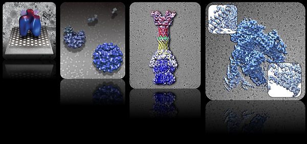
In this presentation, examples where 3D cryo-EM has been successfully applied in order to understand several biological systems will be described. The studies include protein scaffold found on immature Poxvirus particles, retrovirus capsid assembly, multidrug efflux complex of Gram-negative bacteria, and archaeal RNA polymerase. Also, future prospective of constantly evolving 3DEM field will be discussed, with an anticipation of great biological discoveries that were once considered impossible.
Subscribe to the OIST Calendar: Right-click to download, then open in your calendar application.


