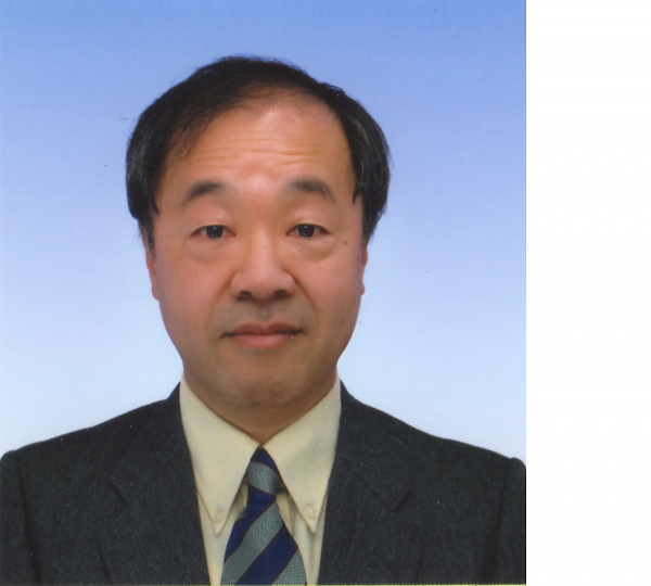
"Functional architecture of cortical microcircuit" Prof. Yoshiyuki Kubota

Date
Location
Description
Prof. Yoshiyuki Kubota
Division of Cerebral Circuitry
National Institute for Physiological Sciences, Okazaki, Japan
Abstract
The neuronal microcircuit of the neocortex is very complex, composed of excitatory neurons including pyramidal neurons (PNs) and spiny stellate cells, inhibitory non-pyramidal interneurons (NPs), glutamatergic afferent axons arising from the other cortical areas as well as subcortical structures including the thalamus together with non-glutamatergic neuromodulatory afferents from many different nuclei. Perturbed activity in the cortical microcircuit is associated with pathologies including epilepsy, autism and schizophrenia. However, mechanisms controlling microcircuit functions are incompletely understood.
In order to understand the functional architecture of the cortical microcircuit I began by classifying the cortical NP subtypes. Distinct patterns of expression of various neurochemical markers including parvalbumin, calretinin, somatostatin, VIP and unique firing properties were observed in various cortical NP subtypes. Next, the inhibitory synaptic connections onto the excitatory PNs were analyzed using combined physiological, morphological and simulation analyses. We found that the cortical NPs are innervated by inhibitory synapses on various subdomains: axon initial segment, soma, dendritic shaft and spine, and the IPSP size induced by these inputs is not uniform. Interestingly the spines receiving inhibitory synapses were shown to also receive a thalamo-cortical excitatory synapse, so these spines were called dually innervated spines (DiS). The inhibitory synapse should efficiently veto the thalamic excitatory input at the DiS. More recently, we have begun to analyze cortical synaptic structural dynamics using two-photon microscopy to visualize fluorescently labeled dendritic spines in the brain of transgenic mice (YFP-H line) during motor skill learning and have identified newly formed synaptic contacts using 3D reconstructions from serial electron micrographs obtained by ATUMtome/SEM or FIB/SEM. We seek to identify the structural and functional basis of newly formed memory traces in the cortical microcircuit. These studies significantly deepen our understanding of the organization of the neuronal circuit and its role in information processing and memory encoding in the mammalian brain.
Biography
Research topic: The Organization of the Cortical Microcircuit
Research techniques: neuroanatomy, EM 3D reconstruction, in vivo imaging, electrophysiology
Current position: Associate Professor, Division of Cerebral Circuitry, National Institute for Physiological Sciences, Okazaki, Japan
Degree: Ph.D. in Medical Sciences, Osaka University, Japan (1988/3)
Professional History:
Associate Professor, Division of Cerebral Circuitry, National Institute for Physiological Sciences, Okazaki, Japan (2001/10-present)
Researcher, Bio-Mimetic Control Research Center, RIKEN, Nagoya, Japan (1994/4-2001/9)
Basic Science Researcher, Frontier Research System, RIKEN, Wako, Japan (1991/4-1994/3)
Postdoctoral Research Fellow, University of British Columbia, Vancouver, Canada (1990/11-1991/3)
Assistant Professor, Kagawa Medical School, Takamatsu, Japan (1990/1-1991/3)
Postdoctoral Research Fellow, University of Tennessee, Memphis, USA (1989/1-12)
Postdoctoral Research Fellowship of JSPS, Osaka University, Osaka, Japan (1987/4-1988/12)
Member:
The Japan Neuroscience Society (1991-), Physiological Society of Japan (2007-), The Japanese Association of Anatomists (2007-), The Society for Neuroscience (1989-)
Subscribe to the OIST Calendar: Right-click to download, then open in your calendar application.


