

OIST-Kyoto University Joint Workshop -Challenges in Biomedical Complexity-
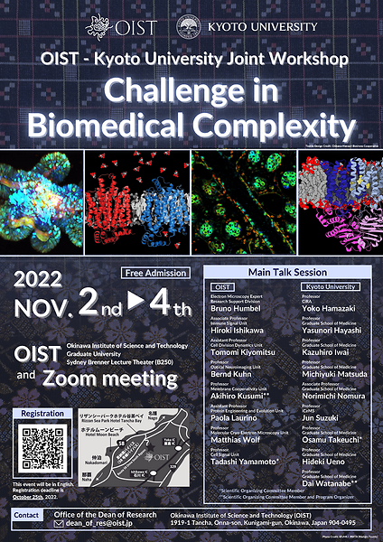
Date
Location
Description
Registration
-Registration is now closed-
Title:
OIST-Kyoto University Joint Workshop -Challenges in Biomedical Complexity-
Symposium Abstract:
OIST and the Kyoto University Graduate School of Medicine are forcibly advancing our frontiers for understanding the biomedical complexities characterized by volatility, uncertainty, and ambiguity, covering from the molecular systems to the entire body. This year's joint seminar features the new important findings made in immunological and brain systems by the scientists at OIST and Kyoto University Graduate School of Medicine, as well as the imaging technologies developed for clarifying the complex systems from molecules to human brain.
Symposium Photos
Click Here
Main Talk Session
| Speakers | Affiliation | Talk Title/Abstract | |
| 1 |
Dr. Tomomi Kiyomitsu, Assistant Professor |
Plasticity of chromosome segregation mechanism during development A fertilized egg undergoes repeated cell division to build a complicated body during development of multi-cellular organisms. In each cell division process, duplicated chromosomes are properly segregated into daughter cells by a microtubule-based structure called the mitotic spindle. Prior studies established a universal model of the mitotic spindle assembly in animal cells, but it is unclear whether and how the mechanism is adapted to diverse contexts during development. In this talk, I will present our recent studies indicating that mechanisms of mitotic spindle assembly and chromosome segregation are unexpectedly different between early embryonic and somatic divisions in vertebrates. I will also highlight two powerful technologies, auxin-inducible degron and light-inducible manipulation in living cells. |
|
| 2 |
Dr. Kazuhiro Iwai, Professor |
Unexpected regulation and function of LUBAC-mediated linear ubiquitination in immunity Linear (M1-linked) ubiquitin chains, which are specifically generated by the LUBAC ubiquitin ligase complex, played crucial roles in inflammatory signaling by inducing NF-kappaB activation and suppressing programmed cell death including apoptosis and necroptosis. LUBAC is composed of three subunits and the HOIP subunit has the catalytic center for linear ubiquitination. The HOIL-1L subunit also has the E3 ligase center and we realized that slower migrating HOIL-1L is disappears by loss of HOIL-1L E3. Our detailed analyses revealed that HOIL-1L conjugates mono-ubiquitin to all three LUBAC subunits and HOIP RBR further conjugates linear ubiquitin chains onto the mono-ubiquitin moieties, which leads to the suppression of linear ubiquitination activity of LUBAC. We further showed that OTULIN, the linear-specific deubiquitinase counteracts suppression of LUBAC function by cleaving linear chains conjugated by HOIL-1L and HOIP coordinately from the LUBAC complex. Introduction of E3-defective HOIL-1L mutants augments NF-kappaB activation and suppresses programmed cell death by enhancing linear ubiquitination of target proteins. We are now dissected roles of linear ubiquitination in inflammatory diseases. Our current progress will be presented in this symposium. |
|
| 3 |
Dr. Paola Laurino, Assistant Professor |
Enzymes’ behavior in highly crowded membrane-less compartments Enzyme-catalysed reactions happen every second in the complex crowded environment of cellular cytosol. Here, we introduce a model system to mimic the high macromolecular crowding found in the cellular cytosol. Our system takes advantage of the tendency of proteins to phase-separate in membrane-less compartments, the lack of membrane barrier allows substrates and products to freely diffuse in the system. We demonstrate how this system can self-generate in situ a pH gradient and in general be used to study enzyme behavior in a macromolecular crowded and controlled environment. Ultimately our system recapitulates putative biological and pre-biotic conditions. |
|
| 4 |
Dr. Jun Suzuki, Professor |
Kyoto University, Institute for Integrated Cell-Material Sciences (iCeMS) |
Unbiased screening approaches to reveal mechanisms of lipid dynamics Cells establish the asymmetrical distribution of molecules across membranes using ATP as an energy and quickly alter this asymmetrical distribution to adopt to the environmental changes. In the case of ions, sodium and calcium ions are exported to the extracellular space, and this electrochemical gradient is utilized to activate cells. In the case of lipids, the phospholipid phosphatidylserine (PS) is distributed restrictively to the inner side of the plasma membrane in living cells while is exposed on the cell surface when cells are activated or undergo cell death. The exposed PS is used for the scaffold for chemical reaction or signal for removal of dead cells and unwanted compartments in living cells. Proteins altering this asymmetrical distribution of lipids are called scramblases, but their molecular identity had been unknown for decades. Using unbiased screening approaches, we identified the long-sought after scramblase(s), their chaperones, activators, and regulators. Based on the analysis of identified genes, their physiological significance was also investigated. In this symposium, I am going to introduce how we approached to identify the molecular components of lipid scrambling and seek the potential collaboration opportunity with OIST researchers. |
| 5 |
Dr. Osamu Takeuchi, Professor |
Post-transcriptional control of immune responses by Regnase-1 and its role in health and disease Post-transcriptional regulation mediated by RNA-binding proteins is critical for the maintenance of innate and adaptive immune responses. We discovered that an RNase Regnase-1 degrades mRNAs encoding immune activating proteins such as inflammatory cytokines by recognizing stem-loop structures present in the 3’ untranslated region (UTR). Regnase-1-mediated mRNA decay requires protein translation and a helicase UPF1. Regnase-1 is critical for the prevention of autoimmunity not only in mice, but also in various human diseases including ulcerative colitis, lung fibrosis, multiple sclerosis and pulmonary hypertension. We recently developed a therapeutic strategy to suppress inflammatory responses by blocking Regnase-1 self-regulation. Regnase-1-targeting antisense oligonucleotides not only enhanced Regnase-1 expression but also effectively reduced the expression of multiple proinflammatory transcripts, and ameliorated acute and chronic inflammation mouse models. In this talk, I will discuss the roles of Regnase-1 in controlling human and mouse immune diseases. |
|
| 6 |
Dr. Tadashi Yamamoto, Professor |
Post-transcriptinal regulation by CCR4-NOT: Lessons from immune cells mRNA decay is a post-transcriptional mechanism controlling mRNA abundance. The carbon catabolite repression 4 (CCR4)-negative on TATA-less (NOT) complex controls mRNA longevity by catalyzing mRNA deadenylation. mRNA decay, regulated by the CCR4-NOT complex, is required for differentiation of pro-B to pre-B cells. In this process, it is likely that the RNA-binding proteins, ZFP36 ring finger protein like 1 and 2, recruit the CCR4-NOT complex to specific target mRNAs, thereby inducing cell quiescence of pro-B cells. The CCR4-NOT complex also participates in positive selection of thymocytes. Mechanistically, the CCR4-NOT deadenylase complex inhibits abnormal apoptosis by reducing the expression level of mRNAs encoding pro-apoptotic proteins, which are otherwise up-regulated during positive selection. |
|
| 7 |
Dr. Hideki Ueno, Professor |
Human Immunology 2.0 The immune system is involved in a much wider variety of disease pathogenesis than was previously anticipated. Whereas mouse models have played an essential role in increasing our knowledge of immunology during the past decades, scientists have realized the limitations in translation to humans. Human immunology using human specimens is vital for understanding immune mechanisms and pathogenesis in humans, and its importance will continue to increase. The technology and tools for human immunology research have advanced dramatically, allowing us to obtain vast amounts of information from even small samples. Immune cells are present not only in blood and lymphoid tissues but also in many organs and play a central role in regulating immune responses in situ. My lab is conducting human immunology research by using blood samples as well as tissue-derived immune cells, including human liver. In my seminar, I would like to introduce several ongoing research on COVID-19, cancer, and liver diseases. |
|
| 8 |
Dr. Hiroki Ishikawa, Associate Professor |
Regulation of AP-1 transcription factors in autoimmune diseases and vaccine responses Host cellular and gut microbial metabolism is closely associated with a variety of immune responses, but how they regulate immune transcriptional programs remains largely unknown. Here, I present our recent findings that indicate novel metabolic regulation of the AP-1-dependent transcriptional program, which plays crucial roles in innate and adaptive immunity. First, I will focus on the mechanism by which the cellular metabolism regulates AP-1-dependent autoimmune T helper cell differentiation. Next, I will report that a specific gut microbial metabolic pathway is associated with the AP-1-mediated transcriptional program that negatively regulates COVID-19 vaccine-induced T cell responses. |
|
| 9 |
Dr. Yoko Hamazaki, Professor |
Kyoto University, Center for iPS Cell Research and Application (CiRA) |
Age-related and individual heterogeneity of immune responses to SARS-CoV-2 mRNA vaccine Advanced age is a significant risk factor for severe symptoms and fatality in patients with infectious diseases, including in those with coronavirus disease 2019 (COVID-19). This is likely because of the age-associated decline in immune competence. Thus, older individuals are strongly recommended to receive vaccines. However, the benefits and efficacy of vaccination in older adults are generally limited, mainly because of the decreased effectiveness of acquired immunity. T cells play a central role in regulating antigen-specific antibodies and cytotoxic responses, but the composition and functionality of T cells greatly change with age. In this presentation, I will introduce our study on how age-related defects in T cells impact vaccine responses by comparing the immunogenicity and reactogenicity of two doses of SARS-CoV2 mRNA vaccine between adults (aged <65 years) and older adults (aged ≥65 years) in a cohort of 216 individuals. Our findings improve the understanding of age-related defects in antigen-specific T-cell responses in humans and will contribute to future vaccine strategies and various immunotherapies, particularly for older populations exhibiting immunosenescence. |
| 10 |
Dr. Michiyuki Matsuda, Professor |
Intercellular communication by growth factors and prostaglandin E2 Paracrine signals mediated by peptides and lipids are the major intercellular communication tools. However, many of the precedent studies adopted the bath application of paracrine factors to tissue culture cells, leaving the effect of endogenous paracrine factors elusive. In fact, the paracrine signaling triggered by the endogenous ligands has rarely been visualized. By using cells expressing biosensors for ERK MAP kinase and protein kinase A (PKA), we have developed systems to visualize paracrine signals mediated by receptor type-tyrosine kinases and Gs protein-coupled receptors (GsPCR). We found that in confluent epithelial cells mechanical stretch triggers ADAM17 activation, which cleaves off ligands for the epidermal growth factor receptor (EGFR). The activated EGFR then stimulates ERK, which, in turn, activates ADAM17, thereby propagating ERK activation waves to the remote cells. Meanwhile, we observed that stochastic calcium transients trigger the switch-like discharge of prostaglandin E2 (PGE2), which activates PKA in neighboring cells via GsPCR. A stark difference between these two systems is in the velocity of signal propagation. While ERK activation propagates at 1~4 µm/min, the PKA activation propagates 100 µm/min within confluent epithelial cells. These observations have delineated the temporal difference of paracrine siganls mediated by growth factors and PGE2. |
|
| 11 |
Dr. Bruno Humbel, Electron Microscopy Expert |
Electron Microscopy in Uchinaa: Reality and Dreams The last couple of years we concentrate on gaining 3D structure information of biological samples using electron microscopy. In the first place we are unraveling the atomic structure of proteins. This is a big national project initiated by AMED’s Basis to Supporting Innovative Drug Discovery and Life Science Research (BINDS). In collaboration with Prof. Tani from Mie University we are analysing light harvesting complexes of different phototrophic bacteria. |
|
| 12 |
Dr. Dai Watanabe, Professor |
Imaging the neural basis of cognitive flexibility in decision making Cognitive flexibility is a fundamental brain function in decision-making that enables us to properly change behaviors in response to environmental changes or risks. Accumulating evidence suggests that the frontal cortex plays critical roles in cognitive flexibility. However, the underlying cellular and circuit mechanisms are not well understood. We developed two different reversal-learning tasks, which are designed to assess cognitive flexibility in prediction-based and non-prediction-based decision-making, respectively. We applied live-cell Ca2+ imaging to study single-unit activity of neurons in the frontal cortex during the tasks, and found that the premotor cortex (M2) and orbitofrontal cortex (OFC) are essential for cognitive flexibility in these tasks, respectively. M2 layer 5 neurons compute the value of behaviors by integrating two different types of information, namely the action-outcome history and the knowledge of the environment, and are required for behavioral change based on prediction of future environmental changes. In contrast, when facing unpredictable changes in environment, OFC layer 2/3 neurons facilitate switching between different behavioral strategies, which is often observed in heuristics-based or intuition-based decision-making in humans. These results suggest that subregions in the frontal cortex differentially contribute to cognitive flexibility depending on complexity of the environment. |
|
| 13 |
Dr. Akihiro Kusumi, Professor |
Development of ultrafast single molecule imaging, PALM, and dSTORM and their appliation to reveal molecular architecture and dynamics of the focal adhesion We developed an ultrafast camera system that enables the highest time resolutions in single fluorescent-molecule imaging, PALM, and dSTORM to date. Their temporal resolutions are photon-limited by fluorophore photophysics: 33 (100) µs with a single-molecule localization precision of 34 (20) nm for Cy3, the optimal fluorophore we found, and 10 s (for 10,000 frames) for PALM/dSTORM imaging of an entire live cell. This camera detected fast hop diffusion of membrane molecules in the plasma membrane (PM) and the fluid membrane part in the focal adhesion (FA). Ultrafast simultaneous two-color PALM/dSTORM revealed that the FA consists of FA-protein islands of ≈50 nm in diameter, which form loose clusters with diameters of ≈350 nm as functional units. The developed ultrafast camera will help to elucidate the principles governing the PM organization. |
|
| 14 |
Dr. Matthias Wolf, Professor |
Assembly mechanism of the pleomorphic immature poxvirus scaffold n Vaccinia virus (VACV), the prototype poxvirus, scaffold protein D13 forms a honeycomb-like lattice on the viral membrane that results in formation of the pleomorphic immature virion (IV). The structure of D13 is similar to those of major capsid proteins that readily form icosahedral capsids in nucleocytoplasmic large DNA viruses (NCLDVs). However, the detailed assembly mechanism of the nonicosahedral poxvirus scaffold has never been understood. Here we show the cryo-EM structures of the D13 trimer and scaffold intermediates produced in vitro. The structures reveal that the displacement of the short N-terminal α-helix is critical for initiation of D13 self-assembly. The continuous curvature of the IV is mediated by electrostatic interactions that induce torsion between trimers. The assembly mechanism explains the semiordered capsid-like arrangement of D13 that is distinct from icosahedral NCLDVs. Our structures explain how a single protein can self-assemble into different capsid morphologies and represent a local exception to the universal Caspar-Klug theory of quasi-equivalence. |
|
| 15 |
Dr. Norimichi Nomura, Associate Professor |
Unveiling membrane protein structures with the aid of antibody toolkits Membrane proteins are the principal targets for drug discovery since they modulate myriad critical biological functions. Attempts to explore the fundamental molecular basis that link membrane protein structure to functions have been hindered by a number of seemingly insurmountable technical barriers. To overcome the impediments, we have developed and implemented a new technique based on antibody generation against conformational epitopes in membrane protein surfaces. While such antibodies have demonstrated efficacy as crystallization chaperones, their use in cryo-EM as fiducial markers is especially impactful in structure determination of smaller membrane proteins (< 50kDa) such as medically relevant transporters and receptors. In this talk, a couple of examples for antibody-aided structural study of membrane proteins will be highlighted, including X-ray crystallography of the sugar transporter GLUT5 (Nomura et al., Nature, 2015) and cryo-EM single particle analysis of the hepatitis B virus entry receptor NTCP (Asami et al., Nature, 2022; Park et al., Nature, 2022). |
|
| 16 |
Dr. Bernd Kuhn, Professor |
Imaging neuronal activity with 2-photon microscopy in awake mice 2-photon microscopy allows 3D reconstruction of neurons and imaging of neuronal activity in awake animals. The activity can be imaged from cellular compartments as well as from large populations of neurons. Additionally, 2-photon microscopy can be combined with behavioral experiments. In my talk, I will present the basics of two-photon microscopy and a few examples of calcium and voltage imaging in cerebral and cerebellar cortex of behaving mice. |
|
| 17 |
Dr. Yasunori Hayashi, Professor |
Online and offline LTP during memory consolidation Memories are initially formed in the hippocampus but subsequently transferred to the rest of brain for a long-term storage in a process called "memory consolidation,". The cellular mechanism responsible for it has not been fully elucidated. LTP of synaptic transmission is known as a cellular phenomenon of memory. We developed a technique to detect when and where LTP is occurring by employing SuperNova, a fluorescent protein. Upon illumination, SuperNova releases reactive oxygen, which inactivates the surrounding proteins. We fused SuperNova with cofilin, an actin binding protein specifically accumulates at the synapse after LTP induction. Illumination of cofilin-SuperNova within 20 min but not beyond after the induction erased LTP in vitro. Illumination of the hippocampus immediately after learning as well as during sleep after the learning erased the memories, indicating that two waves of LTP occurred in the hippocampus. In contrast, in the anterior cingulate cortex, a cortical region implicated in the recall of old memory, LTP was induced during sleep the day after learning but not on the same day. This technology will elucidate brain functions involved in memory at the cellular level. |
*In the order of appearance
Young Scientist Short Talk Session
| 1 |
Dr. Taka A. Tsunoyama, Postdoctoral Scholar |
Nanoscale condensed liquid platform on the plasma membrane for signal integration We found a nanometer-scale liquid-like protein platform for integrating the signals downstream from GPI-anchored receptors and receptor-type tyrosine kinases, inducing the non-linearly amplified PLCg-IP3-Ca2+ signal outputs via the mutual activations of focal adhesion kinase and Src-family kinases. The platform employs some of the focal adhesion proteins, including integrin, talin, RIAM, VASP, and zyxin, but is distinct from focal adhesions, and is thus termed iTRVZ. The iTRVZ formation is driven by the protein liquid-liquid phase separation and the interactions with the raft domains in the plasma membrane and cortical actin. iTRVZ non-linearly integrates the two distinctly different receptor signals, and thus works as an AND logic gate and noise filter. Dwell lifetimes of the iTRVZ constituent molecules and signaling molecules recruited to iTRVZ are in the range of 0.1 ~ 2 s, whereas the iTRVZ lifetime is ~10 s, allowing dynamic signal regulation and secure signal switching off. |
|
| 2 |
Dr. Keiko Yasuda, Assistant Professor |
Antiretroviral therapy (ART) has greatly improved the prognosis of human immunodeficiency virus (HIV-1)-infected patients, but requires lifelong continuation of ART. In order to establish therapies to eradicate HIV-1 from the body, it remains important to understand the full context of the host's defensive immune response against HIV-1. To date, approximately 1700 RNA-binding proteins have been identified in humans, but the role of RNA-binding proteins in biological defense as an antiviral response is only partially understood. Based on the expression screening, we found that MEX3B, an RNA-binding protein, strongly suppresses HIV-1 viral replication depending on its RNA-binding ability. We are currently investigating the mechanism by which MEX3B suppresses HIV-1 replication and it turned out that MEX3B binds directly to HIV-1 RNA and suppresses the release of infectious virions by repressing viral protein translation. I would like to discuss our current findings. |
|
| 3 |
Dr. Prashant Jain, Postdoctoral Scholar |
Directed Evolution Approach to Study Complex Kinase Signaling Protein phosphorylation is a highly abundant and physiologically most important post-translational modification performed together by an enzyme kinases and cofactor adenosine triphosphate (ATP). Even after decades of research, the identification of every single target that a kinase can phosphorylate in the living system remains unimaginable. Since the irregular kinase activities and higher phosphorylation levels are mostly associated with the survival of the cancer cells, probing protein phosphorylation reactions in vitro and in vivo and their detection are of immediate interest. Current methods present in the literature have very limited success and most importantly, they do not allow the detection of the phosphorylated substrate in in vivo. We have designed a new method that relies on a directed evolution approach to evolve the kinase of interest such that it is enzymatically active only in the presence of newly designed co-factors. This approach transfers the enzymatic power of kinase into our hands, allowing us to detect the phosphorylated substrate even in in vivo with the help of traditional biorthogonal reactions. We believe that, if successful, this approach can lead to the discovery of new targets crucial for understanding kinase signaling and cancer-related pathologies. |
|
| 4 |
Ms. Rurika Okuda, PhD student |
Kyoto University |
Unbalanced translocation der(1;7)(q10;p10) as a distinct subtype in MDS and related myeloid neoplasms Found in 1.5-6% of patients with myelodysplastic syndromes (MDS) and other myoid neoplasms, der(1;7)(q10;p10) is an unbalanced translocation whose molecular pathogenesis is poorly understood. To elucidate the clinical and molecular features of der(1;7)(q10;p10)(+) myeloid neoplasm cases, we enrolled 148 cases with der(1;7)(q10;p10) and an additional 3,242 myeloid neoplasm cases as controls and analyzed them through targeted-capture sequencing, RNA-sequencing and ATAC-sequencing. der(1;7)(q10;p10) cases presented with a mutation profile that was unique compared to -7/del(7q), +1q and other myeloid neoplasm cases. Frequent mutations were found in RUNX1, EZH2 and ETNK1 for der(1;7)(q10;p10) cases compared to -7/del(7q) cases (OR=5.58, OR=6.52, and OR=6.23, respectively). The prognosis for der(1;7)(q10;p10) MDS cases was better than -7/del(7q) MDS cases (p=0.098), but poorer than +1q MDS cases (p=0.11). RNA-sequencing showed a down-regulation in 895 genes and an up-regulation in 573 genes. GSEA analysis revealed a down-regulation in cell cycle related pathways and TNFα signaling via NFκB pathways. Motif analysis of open-regions identified through ATAC-sequencing showed prominent GATA transcription factor family motifs. Footprint analysis also showed a possibility for GATA binding to the open-regions of chromatin in der(1;7)(q10;p10) MDS cases. Der(1;7)(q10;p10) presented with a unique clinical, genetic, and transcriptomic profile, defining a unique subset of MDS. |
| 5 |
Dr. Haytham Mohamed, Postdoctoral Scholar |
OIST, Cell Signal Unit |
Characterization of a knock-in mouse model of TFG-associated hereditary motor and sensory neuropathy Hereditary Motor and Sensory Neuropathy (HMSN), Okinawa-type, which is also known as HMSN with Proximal Dominant Involvement (HMSN-P) is an autosomal-dominant, adult onset, slowly progressive neurodegenerative disorder. Mutations in the Tropomyosin-receptor kinase fused gene (TFG) have been causally associated with HMSN-P, namely, P285L. TFG is a ubiquitous cytoplasmic protein that has been shown to play an important role in ER-golgi transportation. However, the effect of the TFG mutation on the development and progression of HMSN-P remains unknown. To elucidate the pathophysiological mechanism underlying HMSN-P, we have generated a knock-in mouse model harboring the human Tfg mutation P285L (herein referred to as TfgP282L). Longitudinal behavioral analysis suggest that TfgP282L mice developed motor deficits by 15 weeks of age and sensory neuropathy by around 19 weeks of age and onwards. Furthermore, we found that they exhibited muscle atrophy in examined hind-limb muscles as well as a reduction in compound muscle action potential similar to that of HMSN-P patients. Our current data suggest that our TfgP282L mouse model recapitulates key physiological characteristics of HMSN-P patients. |
| 6 |
Mr. Takuma Asahi , PhD Student |
Kyoto University |
Lineage heterogeneity underlies the multiple origins and functions of type 1 innate lymphoid cells mRNA decay is a post-transcriptional mechanism controlling mRNA abundance. The carbon catabolite repression 4 (CCR4)-negative on TATA-less (NOT) complex controls mRNA longevity by catalyzing mRNA deadenylation. mRNA decay, regulated by the CCR4-NOT complex, is required for differentiation of pro-B to pre-B cells. In this process, it is likely that the RNA-binding proteins, ZFP36 ring finger protein like 1 and 2, recruit the CCR4-NOT complex to specific target mRNAs, thereby inducing cell quiescence of pro-B cells. The CCR4-NOT complex also participates in positive selection of thymocytes. Mechanistically, the CCR4-NOT deadenylase complex inhibits abnormal apoptosis by reducing the expression level of mRNAs encoding pro-apoptotic proteins, which are otherwise up-regulated during positive selection. |
| 7 |
Dr. Rieko Ajima, Staff Scientist |
E3 ubiquitin ligase HUWE1 regulates planar cell polarity through controlling Pk localization Wnt5a is a secreted molecule, which is important for morphogenesis during development in mice through establishment of planar cell polarity (PCP) in several tissue via activation of the Wnt/PCP pathway. However, molecular mechanisms underlay the regulation of the morphogenesis is poorly understood. In order to identify the molecules, which work downstream of core PCP proteins, we conducted Mass-spec analysis of a core PCP protein, Pk2 binding complex upon Wnt5a stimulation. The analysis revealed that Pk2 was recruited to the proteasome machinery in a Wnt5a stimulation-dependent manner, and found to interact with an E3 ubiquitin ligase, HUWE1. Node specific Huwe1 conditional KO (cKO) displayed mis-localization of Pk2 and caused Left-Right axis formation defect. These data suggested that HUWE1 regulate PCP establishment in node through controlling Pk2 localization. |
|
| 8 |
Dr. Yuto Kita , PhD Student |
Kyoto University |
Utilization of multiplexed CRISPR-Cas3 system for multi-exon skipping in DMD patient-derived iPSCss For developing a versatile therapy for Duchenne muscular dystrophy (DMD), skipping multiple exons from exon 45 to 55 is expected as a promising strategy to restore the dystrophin protein in many DMD patients. However, because the exon 44-55 region is spanning 340 kb in the genomic DNA, induction of 340 kb deletion is not readily achievable with the commonly used genome editing tools. |
| 9 |
Dr. Marvin van Toorn, Research Unit Technician |
NuMA deficiency causes micronuclei via checkpoint-insensitive k-fiber minus-end detachment from mitotic spindle poles Micronuclei resulting from improper chromosome segregation are a source of chromosomal instability. To prevent micronuclei formation, bundled kinetochore-microtubules (k-fibers) must be properly connected to all sister chromatids via their plus-ends, whereas their minus-ends must be clustered at the two opposing spindle poles throughout mitosis. However, how this k-fiber minus-end clustering is maintained remains poorly understood. Here, we show that NuMA depletion results in micronuclei formation through k-fiber minus-end detachment from focused spindle poles during metaphase. Importantly, this k-fiber minus-end detachment creates misaligned chromosomes that maintain chromosome biorientation and evade detection by checkpoint surveillance mechanisms, resulting in abnormal lagging chromosomes in anaphase. This mechanism may therefore represent a previously unappreciated source of genomic instability in dividing cells. |
|
| 10 |
Ms. Eriko Deguchi, PhD Student |
Kyoto University |
Extracellular dynamics of EGFR ligands dictates the mode of intercellular communications Epidermal growth factor receptor (EGFR) is the most extensively studied cell surface receptor, due to its canonical roles in cell growth and oncogenesis. Nevertheless, the physiological nature of its seven ligands remains elusive because of their significant functional redundancy. Moreover, most of the previous studies were undertaken by bath application of EGFR ligands, leaving the roles of endogenous EGFR ligands uncertain. Here, we aimed to determine the extracellular dynamics of each EGFR ligand by using novel pro-EGFR ligand probes fused to two fluorescent proteins at the extracellular and intracellular domains. To examine how far each EGFR ligand could send the signal, we set up a drug-inducible EGFR ligands shedding system and cells expressing a FRET biosensor for ERK MAP kinase, the downstream effector. Upon stimulation, radial spread of ERK activation was observed from the EGFR ligand-expressing cells. Surprisingly, we found that low-affinity EGFR ligands such as EREG and AREG propagated the ERK activation wave more efficiently than did the high-affinity ligands such as TGFα and HB-EGF. We will discuss the mechanism underlying this unexpected observation based on our simulation model. |
| 11 |
Dr. Mohamed Eltabbal, PhD student |
Grasping the tongue : Cerebellar control of adaptive tongue kinematics in mice s Precise spatiotemporal control of the tongue is essential in diverse animal behaviors across species such as catching prey and vocalization. Recent studies have started to unravel complex rodent tongue trajectories during licking including cortex-dependent corrective submovements. However, a description of the cerebellar influence on complex tongue kinematics on a millisecond scale is still lacking. Here we present a novel behavioral task that requires head-fixed mice to intercept a laterally approaching food pellet with their tongue. We combined high-speed videography with deep learning methods to retrieve tongue trajectories during interception. In addition, we performed one-photon calcium imaging of populations of Purkinje neurons during behavior. Our results show that mice optimize tongue speed and movement onset time as they learn to intercept moving food pellets at varying approach speeds. We demonstrate via pharmacological inactivation that these adaptive tongue movements are dependent on cerebellar networks in Crus I and II. We consider our behavioral task as a stepping stone to investigating the neural basis of fast adaptive motor kinematics during object interception. |
|
| 12 |
Qianli Zhang, Researcher |
Kyoto University |
Engineered fast-dissociating antibody fragments for multiplexed super-resolution microscopy, IRIS The imaging fidelity of conventional super-resolution microscopy are limited by spatial interference between multiple antibodies in a confined resolved area. IRIS overcomes the problem and achieves high density multiplexed imaging using exchangeable probes that transiently bind to targets. Nevertheless, generation of a fast-dissociating binder for each target has been challenging. Since the repository of antibody is expanding rapidly, we developed a generalizable engineering strategy to generate fast-dissociating recombinant antibodies for IRIS imaging from off-the-shelf antibodies. The strategy could effectively increase the dissociation rate of existing antibodies by orders of magnitude without compromising binding specificity. We successfully generated dozens of validated IRIS probes and demonstrated multiplexed localization of endogenous proteins in small synaptic connections. Our fast-dissociating antibody probes achieved 4-fold higher label density than conventional super-resolution approaches. Thus, IRIS could visualize the feature of synaptic components with higher fidelity. Our mutagenesis strategy will provide extensive applications for existing antibodies developed in pharmaceutical and biological research. |
Archives
Dr. Tomomi Kiyomitsu / OIST Assistant Professor
"Plasticity of chromosome segregation mechanism during development"
Dr. Kazuhiro Iwai / Kyoto U Professor
"Unexpected regulation and function of LUBAC-mediated linear ubiquitination in immunity"
Dr. Paola Laurino / OIST Assistant Professor
"Enzymes’ behavior in highly crowded membrane-less compartments"
Dr. Jun Suzuki / Kyoto U Professor
"Unbiased screening approaches to reveal mechanisms of lipid dynamics"
Young Scientist Short Talk Session 1
Dr. Osamu Takeuchi / Kyoto U Professor
"Post-transcriptional control of immune responses by Regnase-1 and its role in health and disease"
Dr. Tadashi Yamamoto / OIST Professor
"Post-transcriptinal regulation by CCR4-NOT: Lessons from immune cells"
Dr. Hideki Ueno / Kyoto U Professor
"Human Immunology 2.0"
Dr. Hiroki Ishikawa / OIST Associate Professor
"Regulation of AP-1 transcription factors in autoimmune diseases and vaccine responses"
Dr. Yoko Hamazaki / Kyoto U Professor
"Age-related and individual heterogeneity of immune responses to SARS-CoV-2 mRNA vaccine"
Dr. Michiyuki Matsuda / Kyoto U Professor
"Intercellular communication by growth factors and prostaglandin E2"
Dr. Bruno Humbel / OIST Electron Microscopy Expert
"Electron Microscopy in Uchinaa: Reality and Dreams"
Dr. Dai Watanabe / Kyoto U Professor
"Imaging the neural basis of cognitive flexibility in decision making"
Dr. Akihiro Kusumi / OIST Professor
"Development of ultrafast single molecule imaging, PALM, and dSTORM and their appliation to reveal molecular architecture and dynamics of the focal adhesion"
Young Scientist Short Talk Session 2
Dr. Matthias Wolf / OIST Professor
"Assembly mechanism of the pleomorphic immature poxvirus scaffold"
Dr. Norimichi Nomura / Kyoto U Associate Professor
"Unveiling membrane protein structures with the aid of antibody toolkits"
Dr. Bernd Kuhn / OIST Professor
"Imaging neuronal activity with 2-photon microscopy in awake mice"
Dr. Yasunori Hayashi / Kyoto U Professor
"Online and offline LTP during memory consolidation"
Program
Click Here
Attachments
Subscribe to the OIST Calendar: Right-click to download, then open in your calendar application.