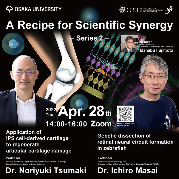
A Recipe for Scientific Synergy Series 2 by Dr. Ichiro Masai and Dr. Noriyuki Tsumaki

Date
Location
Description
Summary:
OIST and Osaka University have been building a healthy relationship.
Speaker, Title and Abstract:
Dr. Ichiro Masai, OIST
Title:"Genetic dissection of retinal neural circuit formation in zebrafish "
The vertebrate retina consists of six major classes of neurons, which form the neural circuit responsible for visual transduction. So, the retina provides a good model for studying the mechanism underlying neural circuit formation. We previously performed a large-scale mutagenesis using zebrafish as an animal model and identified more 300 retinal development-defective zebrafish mutants. Here, I will talk about our recent findings on zebrafish strip1 mutants. Strip1 was initially identified as an interacting protein of Striatin. Strip1 and Striatin function as a hub to form a large protein complex called the STRIPAK complex, which modulates several signaling pathways. Zebrafish strip1 mutants show defects in inner synaptic retinal layer formation. This synaptic layer is normally established by three retinal neurons: retinal ganglion cells (RGCs), amacrine cells, and bipolar cells. In zebrafish strip1 mutants, RGCs undergo dramatic cell death soon after their birth through the activation of a pro-apoptotic factor, Jun. Amacrine and bipolar cells subsequently invade the degenerating RGC layer, leading to a disorganized inner synaptic layer. Thus, Strip1 normally suppresses Jun-mediated apoptotic pathway in RGCs to ensure the formation of synaptic retinal layers. Interestingly, Strip1-deficinet RGCs share similar Jun-mediated stress response with adult mouse and zebrafish RGCs following optic nerve injury. Thus, our findings on the role of Strip1 will advance understanding of pathological process of human glaucoma and hopefully contribute to its therapy development.
Dr. Noriyuki Tsumaki, Osaka University
Title:"Application of iPS cell-derived cartilage to regenerate articular cartilage damage"
Articular cartilage covers the ends of bones and composes joints, providing lubrication between opposing bones during joint motion. Cartilage consists of chondrocytes embedded in abundant extracellular matrix (ECM). Chondrocytes produce and maintain ECM, and ECM is necessary for the chondrocytes to sustain their chondrocytic property including the production of cartilage ECM. Articular cartilage, when damaged through trauma, has only limited capacity for repair, probably because the damage causes a loss of cartilage ECM, disrupting the chondrocytic environment. The continued use of joints with damaged cartilage and poor repair capacity gradually expands the damaged area on the joint surface, resulting in debilitating conditions such as osteoarthritis.
Human induced pluripotent stem cells (hiPSCs) are reprogrammed somatic cells that have pluripotency and self-renew capabilities. We have developed a method by which human iPSCs (hiPSCs) are differentiated toward chondrocytes that produce ECM to prepare cartilage (hiPSC-derived cartilage). We are studying the use of hiPSC-derived cartilage as a curative material to be transplanted into the defect of articular cartilage. To reduce the cost of this regenerative medicine, allogeneic transplantation is preferable. hiPSC-derived cartilage shows low immunogenicity like native cartilage, because the cartilage is avascular and chondrocytes are segregated by the extracellular matrix. After performing pre-clinical tests by transplanting iPSC-derived cartilage into defects created in the articular cartilage of model animals, a clinical test is being implemented.
Moderator:
Dr. Manabu Fujimoto, Osaka University
Zoom:
- https://oist.zoom.us/j/99390867075?pwd=Wkdia1B2R2Vrb0p6ODdVK2lQQTU3dz09
- Meeting ID: 993 9086 7075
- Passcode: 381581
Attachments
Subscribe to the OIST Calendar: Right-click to download, then open in your calendar application.


