FY2020 Annual Report
Membrane Cooperativity Unit
Professor Akihiro Kusumi

Abstract
We at the Membrane Cooperativity Unit are working hard to reveal how the dynamic platforms for signal transduction and the synapses for the neuronal transmission form and function in the plasma membrane. For this purpose, we take a unique approach (in addition to other more conventional approaches). Namely, we develop new and unique methods for single-molecule imaging and manipulation at nanometer precisions in living cells, with a special attention paid to high time resolutions (world’s fastest single fluorescent-molecule imaging). The smooth liaison between physics/engineering and biomedicine is a key for our research.
The plasma membrane is the outermost membrane of the cell, and thus it encloses the entire cell. It is critically important for the cell - the fundamental unit of life - because it defines the space for it. The plasma membrane exchanges information, energy, and substances with the outside world, and we pay special attention to the mechanism for signal transfer from outside to inside the cell, a function generally called “signal transduction”. In the signal transduction process, the plasma membrane works like a sensor + computer + effector.
The Membrane Cooperativity Unit strives to understand how the plasma membrane works at very fundamental levels, based on unique insights we obtain by applying single-molecule imaging-tracking methods. More specifically, we are now revealing the mechanisms by which the metastable molecular complexes and meso-scale membrane domains, including membrane compartments, raft domains, and protein oligomers, form and work in concert to enable signal transduction and synapse formation/modulation in/on the plasma membrane.
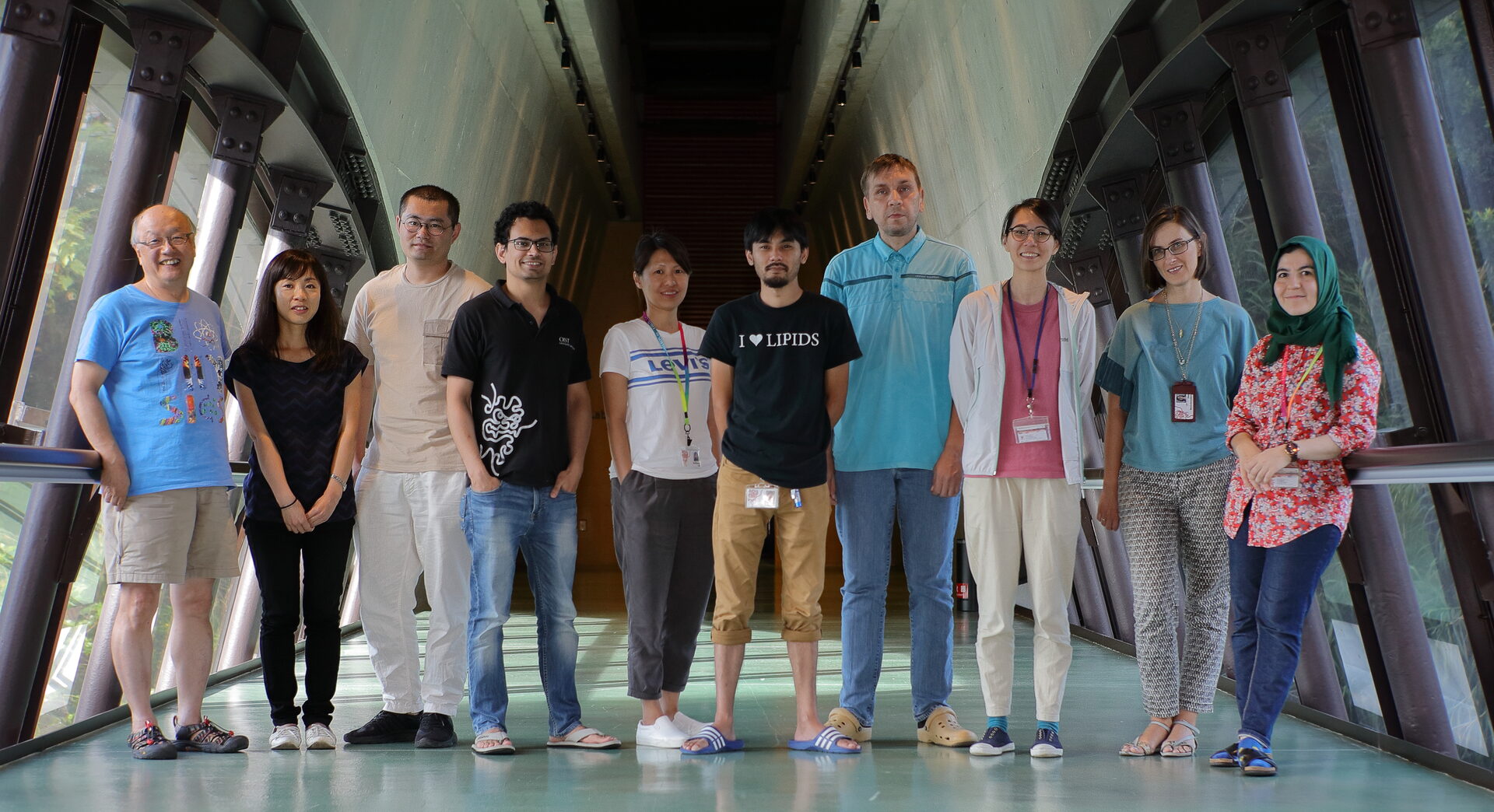
1. Staff
- Dr. Amine Betul Nuriseria Aladag, Post Doctoral Scholar
- Dr. HooiCheng Lim, Post Doctoral Scholar
- Dr. Taka-Aki Tsunoyama, Post Doctoral Scholar
- Dr. Peng Zhou, Post Doctoral Scholar
- Dr. Tim Yeh, Visiting Professor (on sabbatical leave from the University of Texas at Austin)
- Dr. Irina Meshcheryakova, Technician
- Ms. Limin Chen, Technician
- Ms. Hiroko Hijikata, Technician
- Ms. Aya Nakamura, Technician
- Ms. Yuri Nemoto, Technician
- Mr. Yoshifumi Maeda, Research Assistant (Part-time)
- Mr. Tatsuhiro Nishi, Research Assistant (Part-time)
- Mr. Taichi Uchihara, Research Assistant (Part-time)
- Ms. Yuka Nakadomari, Research Assistant (Part-time)
- Mr. Ryoga Maeda, Research Assistant (Part-time)
- Mr. Yuta Kogi, Research Assistant (Part-time)
- Mr. Yusuke Higa, Research Assistant (Part-time)
- Ms. Sachie Matsuoka, Research Unit Administrator (also for Laurino Unit)
- Ms. Miwako Tokuda, Research Unit Administrator
- Dr. Akihiro Kusumi, Professor
2. Collaborations
2.1 Revealing the dynamics, structure, and function of metastable signaling molecular complexes by single-molecule imaging
- Description: Developing ultrafast 3D single-molecule imaging, and applying it to revealing the dynamics and formation mechanism of the signaling complex in synaptic signaling, Fcepsilon signaling, focal adhesion architecture and signaling, and GPI-anchored proteins’ raft-based signaling
- Type of collaboration: Joint research
- Researchers:
- Dr. Takahiro Fujiwara, Associate Professor, Institute for Integrated Cell-Material Sciences (iCeMS), Institute of Advanced Studies, Kyoto University
- Dr. Kenichi Suzuki, Professor, G-CHAIN, Gifu University
2.2 Unraveling the large-scale molecular-species selective diffusion barriers in the axonal initial segment in the neuron using ultrafast single-molecule imaging
- Description: By applying ultrafast single-molecule imaging and ultrafast single-molecule localization microscopy developed by us, we try to unravel the large-scale molecular-species selective diffusion barriers in the axonal initial segment in the neuron
- Type of collaboration: Joint research
- Researchers:
- Dr. Takahiro Fujiwara, Associate Professor, Institute for Integrated Cell-Material Sciences (iCeMS), Institute of Advanced Studies, Kyoto University
2.3 Elucidation of dynamics and formation mechanisms of cellular signaling complexes by developing new single particle tracking methods
- Description: Developing fluorescent probes for their applications to single-molecule imaging in living cells, and by using the developed probes, elucidating dynamics and formation mechanisms of cellular signaling complexes induced by various intercellular signaling molecules and alien antigens, including (non-pathogenic) viruses
- Type of collaboration: Joint research
- Researchers:
- Dr. Dai-Wen Pang, Professor
- Mr. Bo Tang, PhD candidate
- Ms. Dan-dan Fu, PhD candidate
College of Chemistry and Molecular Sciences, Wuhan University, P. R. China
2.4 Revealing the raft-based signaling by using single-molecule imaging
- Description: Revealing the signal transduction mechanisms of GPI-anchored proteins and raft-based signaling by using single-molecule imaging
- Type of collaboration: Joint research
- Researchers:
- Dr. Ikuko Koyama-Honda, Lecturer, Graduate School of Medicine, The University of Tokyo
2.5 Elucidating the functions of plasma membrane compartmentalization
- Description: Elucidating how the signal transduction functions of the plasma membrane is regulated using the actin-based compartmentalization of the plasma membrane, using ultrafast single-molecule imaging-tracking and super-resolution microscopy
- Type of collaboration: Joint research
- Researchers:
- Dr. Pakorn Tony Kanchanawong, Professor, Mechanobiology Institute, The National University of Singapore
2.6 Development of deep-learning methods for single-molecule imaging experiments and analysis
- Description: Developing AI-based methods for performing single-molecule imaging and for analyzing single-molecule imaging data
- Type of collaboration: Joint research
- Researchers:
- Dr. Kazuhiro Hotta, Professor, Mechanobiology Institute, Department of Electrical and Electronic Engineering, Faculty of Engineering, Meijo University
2.7 Revealing the mechanisms for the synapse formation and long-term potentiation by combining super-resolution microscopy and single-molecule imaging
- Description: To discover the mechanisms for functional and structural synaptic plasticity underlying learning and memory, by the combined use of super-resolution microscopy and single-molecule imaging
- Type of collaboration: Joint research
- Researchers:
- Dr. Michisuke Yuzaki, Professor, Graduate School of Medicine, Keio University
3. Activities and Findings
3.1 The signal transduction of the engaged GPI-anchored receptors is critically mediated by the induced transbilayer raft phases: discovery by high-speed single-molecule imaging.
Using single-molecule imaging with enhanced time resolutions down to 5 ms, we found that the CD59-cluster rafts and GM1-cluster rafts were stably induced in the outer leaflet of the plasma membrane (PM), which triggered the activation of Lyn, H-Ras, and ERK and continually recruited Lyn and H-Ras right beneath them in the inner leaflet with dwell lifetimes <0.1 s.
The detection was possible due to the enhanced time resolutions employed here. The recruitment depended on the PM cholesterol and saturated alkyl chains of Lyn and H-Ras, whereas it was blocked by the nonraftophilic transmembrane protein moiety and unsaturated alkyl chains linked to the inner-leaflet molecules. Because GM1 cluster rafts recruited Lyn and H-Ras as efficiently as CD59 cluster rafts, and because the protein moieties of Lyn and H-Ras were not required for the recruitment, we conclude that the transbilayer raft phases induced by the outer-leaflet stabilized rafts recruit lipid-anchored signaling molecules by lateral raft–lipid interactions and thus serve as a key signal transduction platform.
3.2 Dynamic actin-mediated nano-scale clustering of CD44 regulates its meso-scale organization at the plasma membrane
Transmembrane adhesion receptors at the cell surface, such as CD44, are often equipped with modules to interact with the extracellular matrix (ECM) and the intracellular cytoskeletal machinery. CD44 has been recently shown to compartmentalize the membrane into domains by acting as membrane pickets, facilitating the function of signaling receptors. While spatial organization and diffusion studies of membrane proteins are usually conducted separately, here we combined observations of organization and diffusion by using high spatio-temporal resolution imaging on living cells to reveal a hierarchical organization of CD44.
CD44 is present in a meso-scale meshwork pattern where it exhibits enhanced confinement and is enriched in nanoclusters of CD44 along its boundaries. This nanoclustering is orchestrated by the underlying cortical actin dynamics. Interaction with actin is mediated by specific segments of the intracellular domain. This influences the organization of the protein at the nano-scale, generating a selective requirement for formin over Arp2/3-based actin-nucleation machinery. The extracellular domain and its interaction with elements of ECM do not influence the meso-scale organization, but may serve to reposition the meshwork with respect to the ECM. Taken together, our results capture the hierarchical nature of CD44 organization at the cell surface, with active cytoskeleton-templated nanoclusters localized to a meso-scale meshwork pattern.
4. Publications
4.1 Journals
ORIGINAL ARTICLES
- I. Koyama-Honda, T. K. Fujiwara, R. S. Kasai, K. G. N. Suzuki, E. Kajikawa, H. Tsuboi, T. A. Tsunoyama, and A. Kusumi. High-speed single-molecule imaging reveals signal transduction by induced transbilayer raft phases. J. Cell Biol. 219 e202006125 (2020). doi: 10.1083/jcb.202006125
- P. Sil, N. Mateos, S. Nath, S. Buschow, C. Manzo, K. G. N. Suzuki, T. K. Fujiwara, A. Kusumi, M. F. Garcia-Parajo, and S. Mayor. Dynamic actin-mediated nano-scale clustering of CD44 regulates its meso-scale organization at the plasma membrane. Mol. Biol. Cell 31, 561-579 (2020). doi: 10.1091/mbc.E18-11-0715
INVITED REVIEW ARTICLE
1. A. Kusumi, T. K. Fujiwara, T. A. Tsunoyama, R. S. Kasai, A.-A. Liu, K. M. Hirosawa, M. Kinoshita, M. Matsumori, N. Komura, H. Ando, and K. G. N. Suzuki. Defining raft domains in the plasma membrane. Traffic 21, 106-137 (2020). (20th Anniversary Issue of Traffic) doi: 10.1111/tra.12718.
4.2 Books and other one-time publications
Nothing to report
4.3 Oral and Poster Presentations
Poster presentation
- Y. L. Nemoto, K. Naito, H. Hijikata, T. A. Tsunoyama, N. Hiramoto-Yamaki, R. S. Kasai, Y. M. Shirai, M. S. Miyahara, T. K. Fujiwara, A. Kusumi. Dynamic regulation of the AMPA receptor number density in the neuronal synapse as revealed by single-molecule imaging. The 58th Annual Meeting of the Biophysical Society of Japan. Gunma, Japan. Sep.16-18, 2020.
5. Intellectual Property Rights and Other Specific Achievements
Nothing to report
6. Meetings and Events
6.1 Seminar: Time-resolved 3D single-molecule tracking in cytosol and new florescence nanobiosensors
- Date: March 15, 2021
- Venue: OIST Campus Center Building C209
- Speaker: Dr. Tim Yeh (University of Texas at Austin)
7. Other
Nothing to report.


