

Seminar by Mr. En Watanabe (University of Otago)

Date
Location
Description
Abstract:
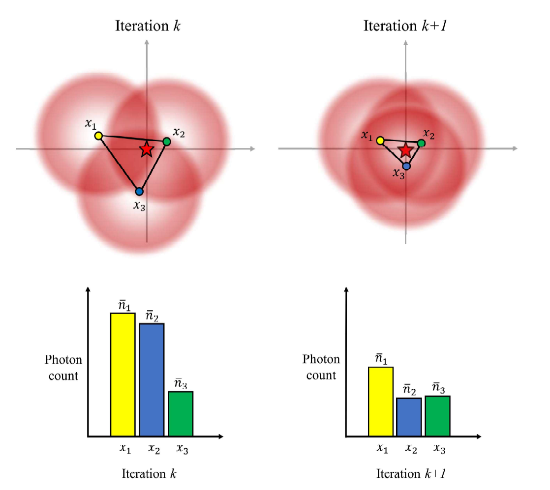
Many techniques have been developed to better understand the cellular processes which govern normal function and pathogenesis in humans. The technique which first allowed scientists to peer into this complex world was light microscopy. However, the nature of light placed a physical limit to the resolution achievable by light microscopy to no less than ∼200 nm. But at the start of the 21st century, techniques which broke this resolution limit emerged, giving rise to the field of super-resolution microscopy (SRM). SRM circumvented the resolution limit through selective or stochastic activation of fluorophores. Initial SRM techniques achieved ∼20 nm resolution, sufficient to resolve some individual large proteins. More recently, a new SRM technique was introduced which achieved ∼1 nm resolution. A low-power excitation beam with a central minimum was employed where localisation of fluorophores was achieved through minimising the recorded photon flux, giving rise to the name of the method: MINFLUX. We aimed to produce an improved MINFLUX microscope and apply it to living biological samples to investigate protein distribution and changes in disease—namely Alzheimer’s disease. This talk will describe our construction process and attempt at MINFLUX microscopy.
Subscribe to the OIST Calendar: Right-click to download, then open in your calendar application.