Publications
Research
What does engram encode?: Heterogeneous memory engrams for different aspects of experience
Nambu MF+, Lin YJ+, Reuschenbach J+, Tanaka KZ*(+:co-first authors)
Curr Opin Neurobiol. June 2022
Long-lasting synaptic changes within the neuronal network mediate memory. Neurons bearing such physical traces of memory (memory engram cells) are often equated with neurons expressing immediate early genes (IEGs) during a specific experience. However, past studies observed the expression of different IEGs in non-overlapping neurons or synaptic plasticity in neurons that do not express a particular IEG. Importantly, recent studies revealed that distinct subsets of neurons expressing different IEGs or even IEG negative-(yet active) neurons support different aspects of memory or computation, suggesting a more complex nature of memory engram cells than previously thought. In this short review, we introduce studies revealing such heterogeneous composition of the memory engram and discuss how the memory system benefits from it.
Characterizing Hippocampal Oscillatory Signatures Underlying Seizures in Temporal Lobe Epilepsy
Mokhothu TM*, Tanaka KZ*(*:co-corresponding authors)
Front. Behav. Neurosci. November 2021
Temporal Lobe Epilepsy (TLE) is a neurological condition characterized by focal brain hyperexcitability, resulting in abnormal neuronal discharge and uncontrollable seizures. The hippocampus, with its inherently highly synchronized firing patterns and relatively high excitability, is prone to epileptic seizures, and it is usually the focus of TLE. Researchers have identified hippocampal high-frequency oscillations (HFOs) as a salient feature in people with TLE and animal models of this disease, arising before or at the onset of the epileptic event. To a certain extent, these pathological HFOs have served as a marker and a potential target for seizure attenuation using electrical or optogenetic interventions. However, many questions remain about whether we can reliably distinguish pathological from non-pathological HFOs and whether they can tell us about the development of the disease. While this would be an arduous task to perform in humans, animal models of TLE provide an excellent opportunity to study the characteristics of HFOs in predicting how epilepsy evolves. This minireview will (1) summarize what we know about the oscillatory disruption in TLE, (2) summarize knowledge about oscillatory changes in the latent period and their role in predicting seizures, and (3) propose future studies essential to uncovering potential treatments based on early detection of pathological HFOs.
An Integrated Index: Engrams, Place Cells, and Hippocampal Memory
Goode TD+, Tanaka KZ+, Sahay A*, McHugh TJ* (+:co-first authors, *:co-corresponding authors)
Neuron August 2020
The hippocampus and its extended network contribute to encoding and recall of episodic experiences. Drawing from recent anatomical, physiological, and behavioral studies, we propose that hippocampal engrams function as indices to mediate memory recall. We broaden this idea to discuss potential relationships between engrams and hippocampal place cells, as well as the molecular, cellular, physiological, and circuit determinants of engrams that permit flexible routing of information to intra- and extrahippocampal circuits for reinstatement, a feature critical to memory indexing. Incorporating indexing into frameworks of memory function opens new avenues of study and even therapies for hippocampal dysfunction.
Heterogeneous representations in the hippocampus
Tanaka KZ*
Neuroscience Research May 2020
The hippocampus is essential for some types of memory, but its specific role remains conjectural. While studies on place cells have supported the hypothesis that the hippocampus provides a spatial substrate for episodic memory, recent engram studies have shown that optogenetic activation of a subset of hippocampal neurons that lack a temporal structure of the spike sequences can also induce memory-associated behavior. In this short review, I discuss the various lines of research that have led to different views of the role of the hippocampus in memory and propose a plausible interpretation of the findings that incorporates two influential theories.
The Hippocampal Engram as a Memory Index
Tanaka KZ*, McHugh TJ
Journal of Experimental Neuroscience Dec 2018
The hippocampus encodes memories for past events, but the nature of the hippocampal code subserving this function remains unclear. A prevailing idea, strongly supported by hippocampal physiology, is the Cognitive Map Theory. In this view, episodic memories are anchored to spatial domains, or allocentric frameworks, of experiences, with the hippocampus providing a stable representation of external space. On the other hand, recent studies using Immediate Early Genes (IEGs) as a proxy of neuronal activation support the Memory Index Theory. This idea posits that the hippocampal memory trace serves as an index for a cortical representation of memory (a map for internal representation) and hypothesizes the primary hippocampal function is to reinstate the pattern of cortical activity present during encoding. Our recent findings provide a unitary view on these two fundamentally different theories. In the hippocampal CA1 region the activity of c-Fos expressing pyramidal neurons reliably reflects the identity of the context the animal is experiencing in an index-like fashion, while spikes from other active pyramidal cells provide spatial information that is stable over a long period of time. These two distinct ensembles of hippocampal neurons suggest heterogeneous roles for subsets of hippocampus neurons in memory.
The hippocampal engram maps experience but not place.
Tanaka KZ*, He H, Tomar A, Niisato K, Huang AJY, McHugh TJ*
Science 361(6400) 392-397 Jul 2018
Episodic memories are encoded by a sparse population of hippocampal neurons. In mice, optogenetic manipulation of this memory engram established that these neurons are indispensable and inducing for memory recall. However, little is known about their in vivo activity or precise role in memory. We found that during memory encoding, only a fraction of CA1 place cells function as engram neurons, distinguished by firing repetitive bursts paced at the theta frequency. During memory recall, these neurons remained highly context specific, yet demonstrated preferential remapping of their place fields. These data demonstrate a dissociation of precise spatial coding and contextual indexing by distinct hippocampal ensembles and suggest that the hippocampal engram serves as an index of memory content.
Single-cell bioluminescence imaging of deep tissue in freely moving animals.
Iwano S, Sugiyama M, Hama H, Watakabe A, Hasegawa N, Kuchimaru T, Tanaka KZ, Takahashi M, Ishida Y, Hata J, Shimozono S, Namiki K, Fukano T, Kiyama M, Okano H, Kizaka-Kondoh S, McHugh TJ, Yamamori T, Hioki H, Maki S, Miyawaki A*
Science 359(6378) 935-939 Feb 2018
Bioluminescence is a natural light source based on luciferase catalysis of its substrate luciferin. We performed directed evolution on firefly luciferase using a red-shifted and highly deliverable luciferin analog to establish AkaBLI, an all-engineered bioluminescence in vivo imaging system. AkaBLI produced emissions in vivo that were brighter by a factor of 100 to 1000 than conventional systems, allowing noninvasive visualization of single cells deep inside freely moving animals. Single tumorigenic cells trapped in the mouse lung vasculature could be visualized. In the mouse brain, genetic labeling with neural activity sensors allowed tracking of small clusters of hippocampal neurons activated by novel environments. In a marmoset, we recorded video-rate bioluminescence from neurons in the striatum, a deep brain area, for more than 1 year. AkaBLI is therefore a bioengineered light source to spur unprecedented scientific, medical, and industrial applications.
Memory retrieval along the proximodistal axis of CA1.
Nakazawa Y, Pevzner A, Tanaka KZ, Wiltgen BJ*
Hippocampus 26(9) 1140-1148 Sep 2016
The proximal and distal segments of CA1 are thought to perform distinct computations. Neurons in proximal CA1 are reciprocally connected with the medial entorhinal cortex (MEC) and exhibit precise spatial firing. In contrast, cells in distal CA1 communicate with the lateral entorhinal cortex (LEC), exhibit more diffuse spatial firing and are affected by the presence of objects in the environment. To determine if these segments make unique contributions to memory retrieval, we examined cellular activity along the proximodistal axis of CA1 using transgenic reporter mice. Neurons tagged during context learning in proximal CA1 were more likely to be reactivated during testing than those in distal CA1. This was true following context fear conditioning and after exposure to a novel environment. Reactivation was also higher in brain regions connected to proximal CA1 (MEC, distal CA3) than those connected to the distal segment (LEC, proximal CA3). To examine contributions to memory retrieval, we performed neurotoxic lesions of proximal or distal CA1 after training. Lesions of the proximal segment significantly impaired memory retrieval while damage to distal CA1 had no effect. These data suggest that context memories are retrieved by a hippocampal microcircuit that involves the proximal but not distal segment of CA1.
Cortical representations are reinstated by the hippocampus during memory retrieval.
Tanaka KZ, Pevzner A, Hamidi AB, Nakazawa Y, Graham J, Wiltgen BJ*
Neuron 84(2) 347-354 Oct 2014
The hippocampus is assumed to retrieve memory by reinstating patterns of cortical activity that were observed during learning. To test this idea, we monitored the activity of individual cortical neurons while simultaneously inactivating the hippocampus. Neurons that were active during context fear conditioning were tagged with the long-lasting fluorescent protein H2B-GFP and the light-activated proton pump ArchT. These proteins allowed us to identify encoding neurons several days after learning and silence them with laser stimulation. When tagged CA1 cells were silenced, we found that memory retrieval was impaired and representations in the cortex (entorhinal, retrosplenial, perirhinal) and the amygdala could not be reactivated. Importantly, hippocampal inactivation did not alter the total amount of activity in most brain regions. Instead, it selectively prevented neurons that were active during learning from being reactivated during retrieval. These data provide functional evidence that the hippocampus reactivates specific memory representations during retrieval.
Systems consolidation and the content of memory.
Wiltgen BJ*, Tanaka KZ
Neurobiology of learning and memory 106 365-371 Nov 2013
Systems consolidation is the process by which memories become independent of the hippocampus and stored in regions of the neocortex. This process is commonly studied in rodents using context fear conditioning. It is becoming increasingly clear, however, that context memories do not always undergo systems consolidation. To explain this fact, the current review describes a number of factors that determine whether or not context fear can be retrieved without the hippocampus during remote memory tests. These include neurogenesis, the presentation of reminder cues after learning, the quality of the memory that is retrieved during testing and the method that is used to inactivate the hippocampus. Based on these data, we propose that remote context fear memories can be retrieved by either the hippocampus or the neocortex. Tests of memory quality (e.g. context discrimination) can typically be used to determine which system is engaged during retrieval. The same is not true of recently formed context fear memories, which appear to always require the hippocampus during retrieval.
Reactivation of neural ensembles during the retrieval of recent and remote memory.
Tayler KK, Tanaka KZ, Reijmers LG, Wiltgen BJ*
Current biology CB 23(2) 99-106 Jan 2013
BACKGROUND:
Episodic memories are encoded within hippocampal and neocortical circuits. Retrieving these memories is assumed to involve reactivation of neural ensembles that were established during learning. Although it has been possible to follow the activity of individual neurons shortly after learning, it has not been possible to examine their activity weeks later during retrieval. We addressed this issue by using a stable form of GFP (H2B-GFP) to permanently tag neurons that are active during contextual fear conditioning.
RESULTS:
H2B-GFP expression in transgenic mice was increased by learning and could be regulated by doxycycline (DOX). Using this system, we found a large network of neurons in the hippocampus, amygdala, and neocortex that were active during context fear conditioning and subsequent memory retrieval 2 days later. Reactivation was contingent on memory retrieval and was not observed when animals were trained and tested in different environments. When memory was retrieved several weeks after learning, reactivation was altered in the hippocampus and amygdala but remained unchanged in the cortex.
CONCLUSIONS:
Retrieving a recently formed context fear memory reactivates neurons in the hippocampus, amygdala, and cortex. Several weeks after learning, the degree of reactivation is altered in hippocampal and amygdala networks but remains stable in the cortex.

Characterization of NMDAR-Independent Learning in the Hippocampus.
Tayler KK, Lowry E, Tanaka KZ, Levy B, Reijmers L, Mayford M, Wiltgen BJ*
Frontiers in behavioral neuroscience 5 28 2011
It is currently thought that memory formation requires the activation of NMDA receptors (NMDARs) in the hippocampus. However, recent studies indicate that these receptors are not necessary for all forms of learning. The current experiments examine this issue using context fear conditioning in mice. First, we show that context fear can be acquired without NMDAR activation in previously trained animals. Mice trained in one environment (context A) are subsequently able to learn about a second environment (context B) in the presence of NMDAR antagonists. Second, we demonstrate that NMDAR-independent learning requires the hippocampus and is dependent on protein synthesis. However, unlike NMDAR-dependent learning, it is not contingent on the expression of activity-regulated cytoskeleton-associated protein (Arc). Lastly, we present data that suggests NMDAR-independent learning is only observed when recently stimulated neurons are reactivated during conditioning. These data suggest that context fear conditioning modifies synaptic plasticity mechanisms in the hippocampus and allows subsequent learning to occur in the absence of NMDAR activation.
Essay
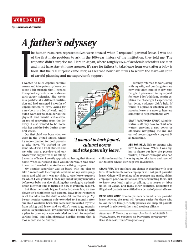
A father's odyssey
Tanaka KZ
Science Vol. 365, Issue 6449, pp. 194 Jul 2019