

FY2014 Annual Report
Brain Mechanism for Behaviour Unit
Professor Gordon Arbuthnott
Abstract
In the fiscal year of 2014, the Brain Mechanisms for Behaviour Unit was made up of six individuals. The latest addition was Yoko Nakano the research technician who has increased the group’s productivity. She has been instrumental in the development of viral vectors for our first steps in optogenetics, and in genotyping the new transgenic animal colonies. We have had a great deal of help from Dr Mike Lazarus and Dr Yoan Cherasse in this development and much of our results have depended on their generous supply of AAV vectors with channel rhodopsin plasmids included. The spring of 2015 Ms. Nakano paid them a visit at the International Institute for Sleep Medicine at the University of Tsukuba to learn how to make AAV viruses including plasmids that express the fluorescent calcium sensor GCaMP6f that she had engineered in OIST.
Dr Violeta Lopez Huerta has been working on the new animals expressing Cre recombinase as well as single cell recording in animals injected with the channel rhodopsin containing viruses. The first of her publications from this work was published electronically in February 2015 but is still not in the paper journal yet. Dr Omar Jaidar has now good evidence that optogenetic stimulation in the striatum in freely moving animals produces dramatic changes in the behavior of mice and is studying the interaction of these effects with dopamine. Dr Takuya Hikima left us in December 2014. This year Dr Marianela Garcia Munoz has some preliminary evidence of behavioral differences within groups of different of transgenic mouse strains and has also completed and published studies of drug effects in cultured cortical and striatal cells. She has contributed strongly to the manuscript writing that has occupied us this year. Three of the 6 manuscripts submitted were published in this fiscal year and two more are in press.
Hermina Nedelescu who initiated studies in the Unit on the neuroanatomy of the cerebellum as part of a scholarship from the Japan Society for the Promotion of Science is now working in Tokyo. This year we had two student interns, Saifullah Khan learned some stereotaxic techniques and joined in some behavioral studies. Johannes Blausenwein under the guidance of Dr. Lopez-Huerta helped in the implementation of the single pellet-reaching task in mice and is acknowledged by authorship in her paper. OIST graduate students on rotation this year were Nilupaer Abudukeyoumu and Jiabao Chen. Ms Abudukeyoumu is now doing her Ph.D. research in the Unit.
1. Staff
- Dr. Marianela Garcia Munoz, Group Leader
- Dr. Takuha Hikima, Researcher (left Dec 2014 and is now at Department of Neurosurgery, NYU-Langone Medical Center, New York, USA)
- Dr. Violeta Lopez Huerta, Researcher
- Dr. Omar Pedro Jaidar Benavides, Researcher
- Ms Yoko Nakano, Technical Staff
- Dr. Hermina Nedelescu, Scholar from the Japan Society for the Promotion of Science (JSPS). Oct 2013 to 2014 now in Tokyo.
- Ms Nilupaer Abudukeyoumu, Graduate Student from April 2014
- Ms Nicola Hazel Guy, MSc student, University of Otago, New Zealand
- Ms Hiroko Chinone, Administrative Assistant
2. Collaborations
- Isolation genetically marked neurons in culture
- Type of collaboration: Joint research
- Researchers:
- Professor William A. Staines, University of Ottawa, Canada
- Professor Antony Krantis, University of Ottawa, Canada
- Dr. Sarah Schock, University of Ottawa, Canada
- Co-ordination of basal ganglia and cerebellar inputs to the motor thalamus.
- Type of collaboration: Joint research
- Researchers:
- Dr. Louise Parr-Brownlie, University of Otago, New Zealand
- Ms. Nicola Hazel Guy, MSc student, University of Otago, New Zealand
- Cortical effects of deep brain stimulation
- Type of collaboration:Joing research
- Researchers:
- Professor Wing Ho Yung, School of Biomedical Sciences, The Chinese University of Hong Kong
3. Activities and Findings
This has been a year of consolidation with 6 papers submitted and 5 in press or published at April 1st 2015. We have continued last year’s projects but serendipity (see section 3.1) required a side project that is already in press.
3.1 Another division of the striatal output neurons
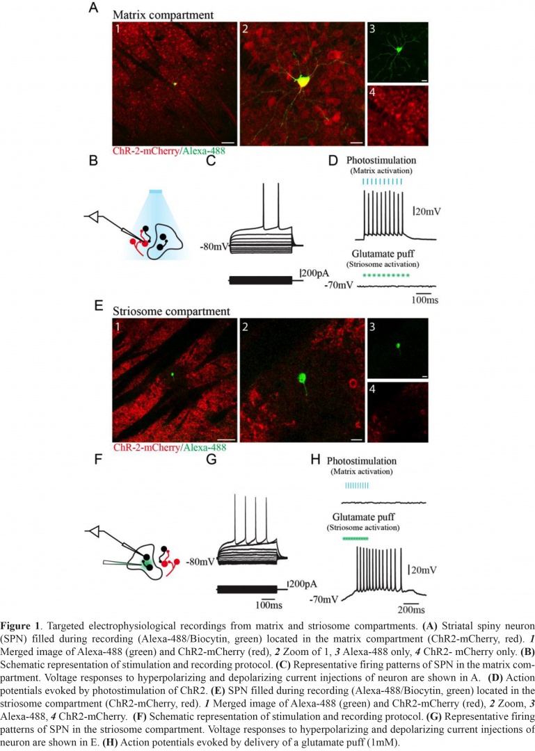
When we first infected striatal output neurons we took care to use an AAV construct that we thought would infect all the cells in the mouse striatum. We checked the outputs to substantia nigra and to globus pallidus and entopeduncular nucleus and all were well stained by the ouput fibres. Such staining assured us that the virus was not biased to either the directly projecting D1 expressing neurons or the indirect pathway extending from D2 neurons. However, when we actualy looked in striatum there were gaps in the staining for the virus. Gaps that looked very like the striosomes, or patches, or dopamine islands on which I had worked in the 1980’s. The different names are the result of this strange compartmental arrangement in the striatum having been described by several people with different methods. Early extensive work from Prof. Ann Graybiel at McGovern Institute for Brain Research at MIT demostrated that all the methods identified the same areas, although with different preferences as to the age of the animals involed. The gaps in the striatal expression of the virus were not stained by calbindin antibodies but were positive for μ-opioid receptor 1 (MOR1) proving that they were indeed the striosomes. Although the early anatomical work suggested that the compartments were separate - neuronal dendrities tended to stay within the borders - no direct evidence of their infividual connectivity had been obtained because of the lack of any in vivo staining method for the two areas. The striosomes were known to form a labyrinth within the matrix of the striatum so sections illustrated them as 'patches'. Not only did our virus localise the matrix cells in vivo and in vitro but the channelrhodopsin with which it infected the cells made them sensitive to light. By recording within a patch and exciting all of the surrounding matrix with light it was clear that none of the strisomal cells responded. They did respond to applied alutamate, but the neighbouring matrix cells did not respond ot their excitation. The two compartments were indeed separate! (See Figure 1).
3.2 Mutual information in cultures.
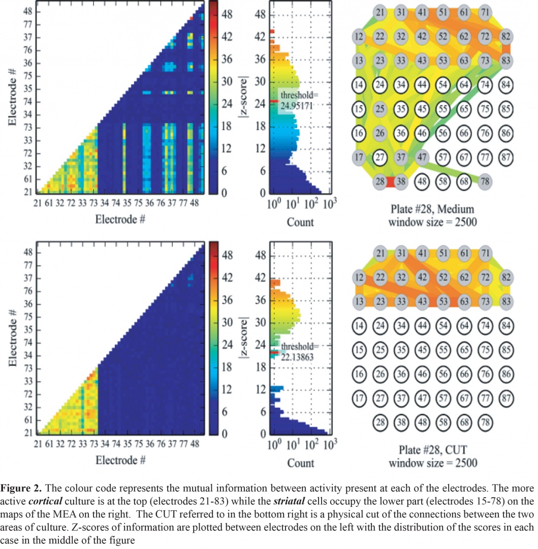
This year also saw the work with the Physics and Biology Unit come to fruition in the publication of a research article in Frontiers in Systems Neuroscience. The re-analysis of experiments done as part of the study of non-synaptic NMDA receptors in striatum (also published this year in Neuropharmacology) let us provide evidence that Mutual Information calculations between two compartments of cultured cells yielded results that were dependent on the integrity of the neuronal connections between the compartments. These experiments supported others that also suggest that connections between cortex and striatum, grown in separate areas of a culture dish, were mainly in one direction – from cortex to striatum. Since this was NOT true in earlier attempts to culture the cells together, it marks a break-through in culturing technology as well as the application of sophisticated mathematical calculations to connectivity in neural networks (see Figure 2).
3.3 How does the brain learn to remember situations and recognize common objects?
One solution to this was proposed in a book published in 1949 by Donald O. Hebb. His idea was that neurons could become connected in assemblies that would fire together when a situation or event happened and with repetition this assembly would come to represent the situation (or object) and as the connections among the assemblies became stronger a small relevant stimulus would ignite the whole assembly and act like a memory of the event or object of importance. He imagined that such assemblies would be formed as the animals became familiar with their world and that cells that usually fire together becoming more strongly connected together would build such assemblies.
Experimentally it was hard to examine the assembly idea because of the need to record enough neurons to include several ‘assemblies’ and ideally with every member present in the recordings. This is becoming possible, and in a recent collaborative work just accepted in International Journal of Neural Systems, we succeeded in finding assemblies, that have all the properties that Hebb described, in groups of cultured cortical neurons whose calcium transients can be recorded with single cell resolution in groups of hundreds of cells at a time. The results are a little surprising because Hebb intended the assemblies to respond to events in the outside world. The cultures have no sensory input from the outside world but are able – indeed seem unable not to - form these assemblies. However, a recent publication by a previous member of the Unit suggests that these assemblies also code for visual information (Carrillo-Reid et al J. Neuroscience 2015).
3.4 Activity of axons in layer 1 of cerebral cortex in awake mice.
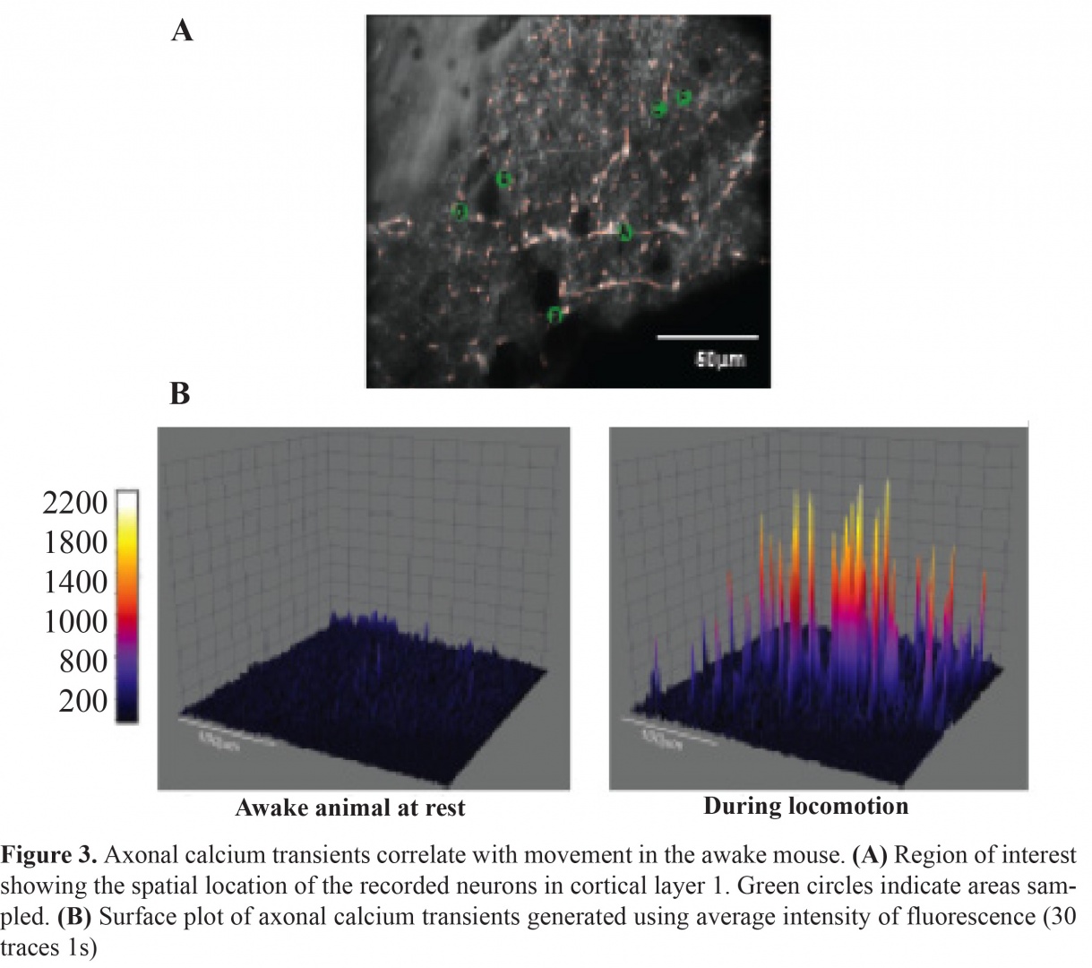
The final output from much of ventral thalamus related to movements is to layer 1 of cortex. Having explored the anatomical distribution of axons from the ventromedial nucleus of thalamus we have now the first images of calcium activity in those same fibers in awake, head fixed, mice in collaboration with Dr Bernd Kuhn head of the Optical Neuroimaging Unit and his team. The difference in activity in mice resting still and moving on a spherical floating ball is dramatic (see Figure 3). Clearly this activity needs to be further analyzed but it already suggests dramatic changes with movement initiation.
3.5 Collaborations within OIST
- Physics and Biology Unit at OIST under the leadership of Professor Jonathan Miller to examine more efficiently the electrical activity in the MEAs, the first result is now published.
- Optical Neuroimaging Unit at OIST under the leadership of Professor Bernd Kuhn to examine, in awake mice, the output pathway from thalamus to cortex that is the final output pathway from the basal ganglia. We have the first images of activity in the thalamocortical fibres in freely moving mice.
- Light matter Interactions Unit we share a Ph.D. student who has been trying to use fiber endoscope technology to image deep in brain tissue. Unfortunately this “New Research Initiative” was not funded and the idea is so popular at present that we may not be the first to succeed, but we stand a good chance of being among the first.
3.6 Collaborations outside OIST
- The collaborative work with several scientists in different countries has been fruitful as well.
- Collaboration with Dr Luoise Parr-Brownlie of the University of Otago, New Zealand, resulted in the arrival in December of Ms Nicola Hazel Guy who is doing research in the Unit towards her master’s degree.
- We continue our collaborative research with Prof. Wing Ho Yung of Hong Kong Chinese University and are working on another publication together.
- Collaboration with Professor William Staines from the University of Ottawa, Canada and Stephan Theiss (Heinrich Heine University of Dusseldorf, Germany) continue as valuable interactions source of interesting and stimulating exchanges.
National collaborations within Japan include:
- Prof Mike Lazarus and Yoan Cherasse at the International Institute for Sleep Medicine at the University of Tsukuba, who have taught us about AAV viruses.
- Dr Tomomi Shimogori at Riken Brain Science Institute who taught us to make embryonic electroporations in mice.
4. Publications
4.1 Journals
- Lopez-Herta, V. G., Nakano, Y., Bausenwein, J., Jaidar-Benavides, O., Lazarus, M., Cherassse, Y., M, G.-M. & Arbuthnott, G. W. The neostriatum: two entities, one structure? Brain Structure and Function, doi:10.1007/s00429-015-1000-4 (2015).
- Garcia-Munoz, M., Lopez-Herta, V. G., Carrillo-Reid, L. & Arbuthnott, G. W. Extrasynaptic glutamate NMDA receptors: Key players in striatal function. Neuropharmacology 89, 54-63, doi:10.1016/j.neuropharm.2014.09.013 (2015).
4.2 Books and other one-time publications
Nothing to report
4.3 Oral and Poster Presentations
- Arbuthnott, G. W. Myelinatedfibres as the source of benefit in deep brain stimulation. 3rd International Conference and Exhibition on Neurology and Therapeutics, Philadelphia, Pennsylvania (2014).
- Arbuthnott, G. W. & Garcia-Munoz, M. TIME FOR A DIFFERENT MODEL OF THE BASAL GANGLIA? Neural Control of Movement Symposium, Amsterdam, The Netherlands (2014).
- Garcia-Munoz, M., Abudukeyoumu, N., Jaidar Benavides, O. P., Lopez-Huerta, V. G., Nakano, Y. & Arbuthnott, G. W. CORTICAL BURSTING ACTIVITY ENHANCED BY BLOCKADE OF ASTROCYTIC GLUTAMATE TRANSPORTER. Biochemical Society and British Neuroscience Society meeting on Astrocytes in neurodegenerative conditions, London, U.K. (2014).
- Hikima, T. & Arbuthnott, G. W. Comparison of the characteristics of two different fluorescent probes for neurotransmitter release. . The 38th annual meeting of the Japan Neuroscience Society, Pacifico Yokohama, Yokohama (2014).
- Hikima, T. & Arbuthnott, G. W. Awakening of presynaptically silent cortical synapses by increasing the size of readily releasable pool. International Society for Neurochemistry Annual Meeting, Yayoi Auditorium, Ichijo Hall/Annex, The University of Tokyo, Tokyo (2014).
- Hikima, T. & Arbuthnott, G. W. Evaluation of presynaptic actions using two different fluorescent probes for neurotransmitter release. Society for Neuroscience 2014, Washington DC, U.S.A. (2014).
- Jaidar, O. P., Luis, C.-R., Lopez-Huerta, V., M, G.-M., Lazarus, M., Hernandez, A., Bargas, J. & Arbuthnott, G. W. Increased activity of direct and indirect striatal pathways underlies the behavioral response to dopamine depletion. NIPS International Workshop and Satellite Symposium of Neuroscience 2014: A Quarter Century after the Direct and Indirect Pathways Model of the Basal Ganglia and Beyond, National Institute for Physiological Sciences, Okazaki, Japan (2014).
- Li, Q., KO, H., CHAN, C. W., Arbuthnott, G. W., KE, Y. & YUNG, W. H. Layer specific inputs to layer V primary motor cortex exhibit different profiles of training-induced synaptic plasticity. Society for Neuroscience 2014, Washington DC, U.S.A. (2014).
- Nedelescu, H., Takeyama, K., Chowdhury, T., Wable, G., Arbuthnott, G. W. & Aoki, C. (In) Activity-induced structural changes of the cerebellar cortex noradrenergic fibers. Society for Neuroscience 2014, Washington DC, U.S.A. (2014).
- Arbuthnott, G. W. Myelinated fibres as the source of benefit in Deep Brain Stimulation., Schoold of Electrical, Electronic and Communications Engineering, University College Dublin (2014).
- Arbuthnott, G. W. Deep brain stimulation: a mysterious success or a clue to help localize the causes of Parkinsonism?, http://hstalks.com/?t=BL1833661-Arbuthnott (2014).
- Arbuthnott, G. W. Time for a different model of the basal ganglia?, Neural Control of Movement Symposium, Amsterdam (2014).
- Arbuthnott, G. W. Facebook for Neuron? Helping isolated neurons from a 'social' network. . OIST Graduate University (2014).
- Arbuthnott, G. W. Time for a new model of the basal ganglia?, Kyusyu University (2015).
- Arbuthnott, G. W. Beware! Models of the basal ganglia go out of date., Hong Kong University of Science and technology, Hong Kong (2015).
- Arbuthnott, G. W. Research in Brain Mechanisms for Behaviour Unit, OIST Graduate University. Kaiho High School (2015).
5. Intellectual Property Rights and Other Specific Achievements
I have been involved in two graduate student courses taught in OIST Neurobiology (B05) April - August 2014, Controversies in Science (A401) September - December 2014.
6. Meetings and Events
6.1 Relation of Vesicle Positioning to Pool Identity and Exocytotic Fusion Mode:Insight from Three-dimensional, Nanometer-Accuracy Tracking of Single Synaptic Vesicles in Hippocampal Neurons.
- Date: October 20, 2014
- Venue: OIST Campus Lab1
- Speaker: Dr Hyokeun Park (Hong Kong University of Science and Technology)
6.2 Mortor Cortex Involvement in Motor Learning Deficits in Parkinsonism.
- Date: October 30, 2014
- Venue: OIST Campus Lab1
- Speaker: Dr. Qian Li (Chinese University of Hong Kong)
6.3 Correcting abnormal motor thalamus activity in parkinsonian rats improves movements.
- Date: December 18, 2014
- Venue: OIST Campus Lab1
- Speaker: Dr. Louise Parr-Brownlie (University of Otago)
6.4 Molecular mechanisms of dentrite patterning---how we can use in utero electroporation and what we have found.
- Date: January 16, 2015
- Venue: OIST Campus Lab1
- Speaker: Dr. Tomomi Shimogori (RIKEN Brain Research Institute)
6.5 Why coffee wakes us up? -- Novel roles of adenosine A2A receptors in sleep-wake regulation.
- Date: March 3, 2015
- Venue: OIST Campus Lab1
- Speaker: Dr. Michael Lazarus (University of Tsukuba)
7. Other
Nothing to report.