

Research
Research Summary
Two of the key questions in neuroscience are how sensory information is represented in the brain and how behavior arises from neuronal activity. To address these questions, the Optical Neuroimaging Unit develops, improves, and applies techniques for imaging neuronal activity in awake, behaving mice. Synthetic and genetically encoded fluorescent probes convert neuronal activity into optical signals and two-photon microscopy allows imaging these optical signals in the brain of awake mice. During imaging, the mice might be exposed to sensory stimuli or perform tasks in a virtual reality system, and thereby allow to study the relation between neuronal activity and sensory stimulation and behavior. Different types of neuronal activity can be studied by using different fluorescent probes.
Calcium Imaging
In collaboration with the Doya Unit, the Kuhn Unit used calcium imaging in populations of neurons to study how the internal model of the external world in a mouse is updated by its own action and sensory cues (Funamizu et al. 2016).
With custom built two-photon microscopes the Optical Neuroimaging Unit also pushes the limits of imaging depth. One major project is to image neuronal calcium activity in the deepest layer of cortex, layer 6, in mice. Calcium transients of layer 6 neurons in visual cortex are observed during different visual stimuli and different behavioral states, from running to sleep (Augustinaite & Kuhn under review). This is important to know because layer 6 is providing feedback in the thalamo-cortical circuit.
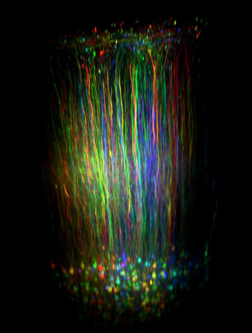
Layer 6 neurons projecting to primary thalamus in visual cortex of the mouse reconstructed in vivo with two photon microscopy. The dendrites of many neurons span the full cortical column, corresponding to about 800 µm. The accompanying manuscript describes the calcium activity of these neurons during different behavioral states. (Augustinaite and Kuhn 2020)
Another important and challenging project is imaging of calcium activity in cortical astrocytes during different behavioral states which reveals a new complexity level of astrocyte signaling. Microdomains can be observed and correlated with the behavioral state of the animal.
Voltage Imaging
A new technology developed in the Optical Neuroimaging Unit is to measure membrane voltage changes optically in single neurons of animals. Now it is possible to simultaneously image dendritic voltage and calcium changes while recording electrical activity from the somata of Purkinje neurons in awake animals. This technique not only allows detecting supra-threshold activity but also sub-threshold activity (Roome & Kuhn 2018). Additionally, the neuronal activity detected is 10-100 times faster than typical calcium imaging data. To do this project, a pure electrochromic voltage-sensitive dye (Kuhn & Fromherz 2003), an optimal imaging technique (Kuhn, Fromherz, Denk 2004), and a chronic cranial window allowing local brain manipulations (Roome & Kuhn 2014) were developed. With this technique it is for the first time possible to observe dendritic information processing in the awake animal on a millisecond time scale. Voltage imaging can also be used to measure brain oscillations. Lately, slow gamma oscillations in cortical layer 1 were discovered with this method (Dalphin et al under review).
Imaging of Protein Kinase A Activity
Additionally, the Optical Neuroimaging Unit images also slow molecular processes in the brain: Protein kinase A activity in somata and dendrites of cortical neurons works changes on a time scale of 10s of seconds in awake, behaving animals. Protein kinase A activity is an important indicator of long-term changes in neurons and it is involved in memory formation.