FY2022
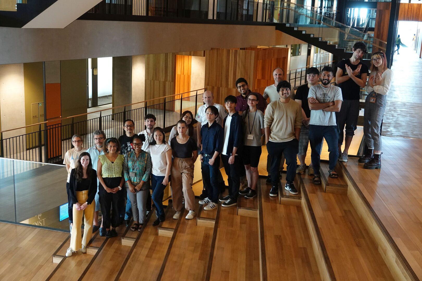
(front, from left to right) Charlotte Denman, Lina Koronfel, Thato Mokhothu, Shinobu Nomura, Aleksandra Gavrilova, Rintaro Shimojo, Kota Shirahata, Amela Londo, Anshuman Nayak, Soumen Jana, Saffira Yan Tjon
Abstract
In FY 2021 the Optical Neuroimaging Unit continued the projects of the previous years. Research projects focused on imaging neuronal and astrocytic activity in the brain of awake mice.
1. Staff
- Dr. Bernd Kuhn, Professor
- Dr. Christopher J. Roome, Staff Scientist
- Dr. Melody Li, Postdoc
- Dr. Sigita Augustinaite, Technician
- Dr. Shinobu Nomura, Technician
- Dr. Mykola "Nik" Medvidov, Technician
- Dr. Kazuo Mori, Technician
- Soumen Jana, Ph.D. Student
- Lina Koronfel, Ph.D. Student
- Mohamed Mostafa Eltabbal, PhD Student
- Cedric Galetzka, PhD Student
- Isabel Anai Echeverria Oviedo , Master Student
- Miles Desforges, PhD Student (co-supervised by Kenji Doya)
- Thato Mokhothu, PhD Student (co-supervised by Kazumasa Tanaka)
- Gaston Sivori, PhD Student (so-supervised by Tomoki Fukai)
- Aleksandra Gavrilova, PhD Student
- Taisuke Higuchi, PhD Student
- Saffira Yon Tjon, PhD Student
- Qiyi Qian, PhD Student
- Amela Londo, Intern Student
- Rintaro Shimojo, Intern Student
- Manami Gima, Research Unit Administrator
Stem Cell and Organoid Research Group
- Dr. Hoang-Dai Tran, Postdoctoral Scholar
- Charlotte Denman, Ph.D. Student
- Runrun Han, Research Technician
2. Collaborations
2.1 High-resolution imaging of barrel cortex through VSD and LFP recordings
- Description: Combining high-resolution electical recording with electrode arrays and voltage imaging
- Type of collaboration: Joint research
- Researchers:
- Associate Professor Stefano Vassanelli, Padua University, Italy
- Dr. Claudia Cecchetto, Junior Researcher, NEU-PAGES Project Team Leader, Padua University, Italy
2.2 Motion correction of in vivo 2P imaging data
- Description: Development of algorythms for motion correction of 2P imaging data
- Type of collaboration: Joint research
- Researcher:
- Philipp Flotho, MSc., Systems Neuroscience & Neurotechnology Unit, Saarland University, Germany
2.3 Development of a genetic targeting mechanism for the pure electrochromic voltage-sensitive dye ANNINE-6
- Description: Synthesis of new voltage-sensitive dyes and in vivo testing of labeling
- Type of collaboration: Joint research
- Researcher:
- Dr. Eugene Khaskin, Science and Technology Group, OIST
2.4 Development of 2D and 3D human pluripotent stem cell differentiation platform for human disease modeling
- Description: Development of 2D and 3D human pluripotent stem cell differentiation platform for human disease modeling
- Researchers:
- Professor Dong Ryul Lee, , CHA University, Korea.
- Dr. Min-Kyoung Shin, CHA University, Korea.
2.5 Neutron-activated, plasmonically excitable Fe-Pt-Yb2O3 core-shell nanoparticles delivering anti-cancer radiation against human glioblastoma brain cancer cells
- Description: Development of novel core-shell nanoparticles which can be neutron-activated, plasmonically excited
- Researcher:
- Dr. Klaus Seemann, Peter Grünberg Institute, Forschungszentrum Jülich, Germany; Université de Lorraine, CNRS, Nancy, France
2.6 Anoxia-Induced LTP at Hippocampal CA1 Synapses
- Description: Imaging astrocytes in vitro during and after anoxia
- Researchers:
- Professor Tomoyuki Takahashi, OIST
- Dr. Han-Ying Wang, OIST
- Isabel Anaí Echeverría Oviedo, PhD Student, NERF in Leuven, Belgium
2.7 Effect of the human FoxP2 gene on a licking task in mouse
- Description: Calcium imaging during a licking task in transgenc mice
- Researchers:
- Adjunct Professor Svante Pääbo, OIST
- Dr. Xiang-Chun Ju, OIST
2.8 TOB is an effector of the hippocampus-mediated acute stress response.
- Description: TOB is an effector of the hippocampus-mediated acute stress response.
- Researchers:
- Dr. Mohieldin Youssef M., OIST
- Professor Tadashi Yamamoto, OIST
3. Activities and Findings
3.1 Voltage Imaging
We improve voltage imaging by synthesizing new dyes and applying them in cerebellar and cerebral cortex. In cerebellar Purkinje neurons, we study dendritic voltage changes during sensory stimulation, the effect of interneurons on dendritic voltage changes, and voltage changes during eye blink condutioning.
3.2 Ca2+ imaging in cortical layer 6
We study neuronal Ca2+ activity in layer 6 of visual cortex during different behavioral states.
3.3 Behavioral state-dependent protein kinase A activity modulated by noradrenaline and dopamine in layer II/III of somatosensory cortex
The catecholaminergic system influences animal behavior by modulating neuronal activity through G-protein coupled receptors and downstream signaling, involving protein kinase A (PKA). Currently, little is known about the real-time dynamics of cortical PKA activity in awake animals. Here, we imaged PKA activity in dendrites and somata of somatosensory cortex layer II/III with two-photon microscopy and the sensor GAkdYmut. Tonic PKA activity was maintained during wakefulness through noradrenaline (NA) and dopamine (DA). Voluntary locomotion induced, on average, biphasic PKA activity changes; decrease during locomotion followed by an overshooting rebound after locomotion offset. Individual somata and dendrites showed a continuum of responses induced by NA and DA. The PKA activity transients of individual dendrites and somata typically changed with every locomotion event, while forced locomotion, associated with stress, induced more cell-specific PKA activity. Positive and negative PKA responses showed a hysteresis, fine-tuning subsequent tonic PKA activity, and thereby excitability and synaptic transmission.
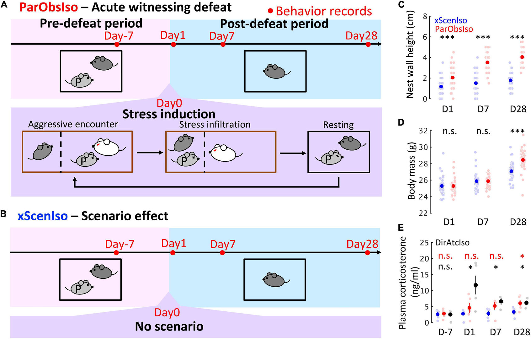
3.5 Social Relationship as a Factor for the Development of Stress Incubation in Adult Mice
While stress reactions can emerge long after the triggering event, it remains elusive how they emerge after a protracted, seemingly stress-free period during which stress incubates. Here, we study the behavioral development in mice isolated after observing an aggressive encounter inflicted upon their pair-housed partners. We developed a spatially resolved fine-scale behavioral analysis and applied it to standard behavioral tests. It reveals that the seemingly sudden behavioral changes developed gradually. These behavioral changes were not observed if the aggressive encounter happened to a stranger mouse, suggesting that social bonding is a prerequisite for stress incubation in this paradigm. This finding was corroborated by hemisphere-specific morphological changes in cortex regions centering at the anterior cingulate cortex, a cognitive and emotional center. Our non-invasive analytical methods to capture informative behavioral details may have applications beyond laboratory animals.
3.6 Ca2+ activity maps of astrocytes tagged by axoastrocytic AAV transfer
Astrocytes exhibit localized Ca2+ microdomain (MD) activity thought to be actively involved in information processing in the brain. However, functional organization of Ca2+ MDs in space and time in relationship to behavior and neuronal activity is poorly understood. Here, we first show that adeno-associated virus (AAV) particles transfer anterogradely from axons to astrocytes. Then, we use this axoastrocytic AAV transfer to express genetically encoded Ca2+ indicators at high-contrast circuit specifically. In combination with two-photon microscopy and unbiased, event-based analysis, we investigated cortical astrocytes embedded in the vibrissal thalamocortical circuit. We found a wide range of Ca2+ MD signals, some of which were ultrafast (≤300 ms). Frequency and size of signals were extensively increased by locomotion but only subtly with sensory stimulation. The overlay of these signals resulted in behavior-dependent maps with characteristic Ca2+ activity hotspots, maybe representing memory engrams. These functional subdomains are stable over days, suggesting subcellular specialization.
Now, we aim to understand the origin of these maps and their involvement in memory.
3.7 Scn2a insufficiency causes altered in vivo neuronal calcium activity and coactivity.
SCN2A protein-truncating variants (PTV) can result in neurological disorders such as autism spectrum disorder and intellectual disability, but they are less likely to cause epilepsy in comparison to missense variants. While in vitro studies showed PTV reduce action potential firing, consequences at in vivo network level remain elusive. Here, we generated a mouse model of Scn2a insufficiency using antisense oligonucleotides (Scn2a ASO mice), which recapitulated key clinical feature of SCN2A PTV disorders. Simultaneous two-photon Ca2+ imaging and electrocorticography (ECoG) in awake mice showed that spontaneous Ca2+ transients, as well as neuronal pairwise co-activities were decreased in Scn2a ASO mice during spontaneous awake state and induced seizure state. The overall reduction in in vivo single neuronal activities and neuronal paired co-activity may likely be the disease mechanisms driving SCN2A ASD and ID.
3.8 Establishment of a new method to create skeletal muscle organoids from human pluripotent stem cells for modeling myogenesis and muscle regeneration
In vitro organoids derived from human pluripotent stem cells (hPSCs) have been developed as essential tools to study the underlying mechanisms of human development and diseases owing to their structural and physiological similarity to corresponding organs. Despite recent advances, there are a few methodologies for three-dimensional (3D) skeletal muscle differentiation, which focus on the terminal differentiation into myofibers and investigate the potential of modeling neuromuscular disorders and muscular dystrophies. However, these methodologies lacked to recapitulate the developmental processes and lacked regenerative capacity. In this study, we developed a new method to differentiate hPSCs into a 3D human skeletal muscle organoid (hSkMO). This organoid model could recapitulate the myogenesis process and possess regenerative capacities of sustainable satellite cells (SCs), which are adult muscle stem/progenitor cells capable of self-renewal and myogenic differentiation. Our 3D model demonstrated myogenesis through the sequential occurrence of multiple myogenic cell types from SCs to myocytes. Notably, we detected quiescent, non-dividing SCs throughout the hSkMO differentiation in long-term culture. They were activated and differentiated to reconstitute muscle tissue upon damage. Thus, hSkMOs can recapitulate human skeletal muscle development and regeneration and may provide a new model for studying human skeletal muscles and related diseases.
3.9 Neutron-activated, plasmonically excitable Fe-Pt-Yb2O3 core-shell nanoparticles delivering anti-cancer radiation against human glioblastoma brain cancer cells
Magnetic nanoparticles can be functionalized in many ways for biomedical applications. Here, we combine four advantageous features in a novel Fe-Pt-Yb2O3 core-shell nanoparticle. (a) The nanoparticles have a size of 10 nm which allow them to diffuse or drift through neuronal tissue as single particles or small clusters. (b) The particles are superparamagnetic after synthesis and ferromagnetic after annealing, enabling directional control by magnetic fields, enhance NMRI contrast, and hyperthermia treatment. (c) After neutron-activation of the shell, they carry low-energetic, short half-lifetime, anti-cancer b-radiation from 175Yb, 177Yb, and 177Lu. (d) Additionally, the particles and their clusters can be optically visualized by plasmonic excitation through multi-photon absorption and the resulting luminescence. To show the potential of the particles for cancer treatment, we exposed cultured cells to non-activated and activated particles, confirm that the particles are internalized, and that the b-radiation of the radioisotopes incorporated in the neutron-activated shell of the nanoparticles kills more than 98% of the LN-18 cancer cells, making the Fe-Pt-Yb2O3 core-shell nanoparticles a promising candidate for future anti-cancer applications.
4. Publications
4.1 Journals
4.2 Books and other one-time publications
Nothing to report.
4.3 Oral and Poster Presentations
- L. Koronfel, C.J. Roome, and B. Kuhn (2022) Dendritic voltage signaling in Cerebellar Purkinje Neurons during Associative Motor Learning. JNS, Okinawa, Japan
- S. Augustinaite and B. Kuhn (2022) Three protocols associated or combinable with long-term two-photon microscopy in head-fixed mice. JNS, Okinawa, Japan
- S. Nomura, P. Flotho, B. Kuhn (2022) PKA activity in layer 2/3 of somatosensory cortex in behaving mice. JNS, Okinawa, Japan
- S. Jana, C.J. Roome, and B. Kuhn (2022) Modulation of Dendritic Voltage Changes in Purkinje Neurons by Inhibitory Inputs in Awake Mice. JNS, Okinawa, Japan
- M. Li and B. Kuhn (2022) Examining the in vivo neural network activity of a Scn2a loss-of-function mouse model. JNS, Okinawa, Japan
- M. Medvidov, C.J. Roome, and B. Kuhn (2022) Application of Expansion Microscopy for 3D Reconstruction of Purkinje Neurons after Functional Imaging in Awake Animals. JNS, Okinawa, Japan
- K. Mori, K. Kurima, J. Wickens, and B. Kuhn (2022) Retro-orbital virus injection allows sparse labelling of neurons for in vivo 2-photon microscopy. JNS, Okinawa, Japan
- C. Cecchetto, B. Kuhn, and S. Vassanelli (2022) Modulation of Dendritic Voltage Changes in Purkinje Neurons by Inhibitory Inputs in Awake Mice. JNS, Okinawa, Japan
- L. Koronfel, C.J. Roome, and B. Kuhn (2022) Dendritic voltage signaling in Cerebellar Purkinje Neurons during Associative Motor Learning. FENS, Paris, France
- S. Nomura, P. Flotho, B. Kuhn (2022) PKA activity in layer 2/3 of somatosensory cortex in behaving mice. FENS, Paris, France
- M. Eltabbal (2022) Grasping the tongue, Cerebellar control of adaptive tongue kinematics. OIST-Kyoto Joint symposium.
- M. Eltabbal (2022) Grasping the tongue, Cerebellar control of adaptive tongue kinematics. JNS Meeting, Okinawa.
4.4 Lectures
- Ajou University – OIST Joint Symposium, Bernd Kuhn (March 7, 2023) Imaging Neuronal Activity with Two-Photon Microscopy in Awake Mice, OIST
- OIST - Kyoto University Joint Workshop: Challenges in Biomedical Complexity. Bernd Kuhn (November 4, 2022) Imaging Neuronal Activity with Two-Photon Microscopy in Awake Mice. OIST
- Fujita Health University. Bernd Kuhn (June 21, 2022) Imaging Neuronal Activity with Two-Photon Microscopy in Awake Mice. Online
5. Intellectual Property Rights and Other Specific Achievements
5.1 JSPS Fellowships
- Mohamed Mostafa Kamal Eltabbal
- Lina Koronfel
- Dr. Melody Li
5.2 Kakenhi
- Christopher J. Roome, Transformative Research Areas (A)
6. Meetings and Events
6.1 Ajou University – OIST Joint Symposium
- Neuroscience and Molecular Medicine
- Date: March 7, 2023
- Venue: OIST
- Organizers: Junghyun Jo (Ajou University) and Bernd Kuhn (OIST)
6.2 ICIIBMS 2022
- 7th International Conference on Intelligent Informatics and BioMedical Sciences (ICIIBMS)
- Date: November 24-26, 2022
- Venue: Nara, Japan
- Organized by researchers of the University of the Ryukyus, the Okinawa National College of Technology, and OIST

7. Other
7.1 Outreach Activities
- A. Gavrilova (2022) Qualitative research methods. Research Workshop for DoDEA High School Teachers, OIST
- A. Gavrilova (2022) Life as a researcher. Yokohama School visit, OIST
- A. Gavrilova (2022) Research and education in Russia and Japan. European Research Days Japan, online
- A. Gavrilova (2022) Life as a researcher. Ibaraki Midori Oka High School visit, OIST.
- K. Mori (2022) Onna-son/OIST Children’s School of Science. Online.
- A. Gavrilova (2022) Neuroscience and Technology. Exchange Program for Asian Students (EPAS), online
- A. Gavrilova (2022) Life as a researcher. Oita Uenogaoka High School visit, OIST.
- A. Gavrilova (2023) Examples of leadership in Neuroscience. Ryukyu University, Okinawa and online.
- K. Mori (2022) Onna-son/OIST Children School of Science. Online.