

Yuki Nakazawa, Ph.D.
Cell biology and morphology
Background
I received my Ph.D. (Science) from the University of Tokyo (2008). My dissertation was about how the 9-fold symmetry of the centriole/basal body and the flagellar axoneme is invariably established. Then, I worked as an assistant professor at Kitasato University, subsequently worked as a research associate at the University of Tokyo. Up to this point, my research focus had been on cell biology, especially the cytoskeleton. In 2012, I joined the group working on memory with my husband at the University of California, Davis. After returning to Japan, I restarted my research about centriole assembly as a JSPS research fellow (RPD), a part-time lecturer, and a research associate at Hosei University (2016-2020). At OIST, I’m continuing this project.
Research interests
I’m studying centriole assembly using green alga Chlamydomonas reinhardtii, a unicellular organism that swims using two flagella (Fig.1).
1) About Chlamydomonas
Chlamydomonas has the following advantages: 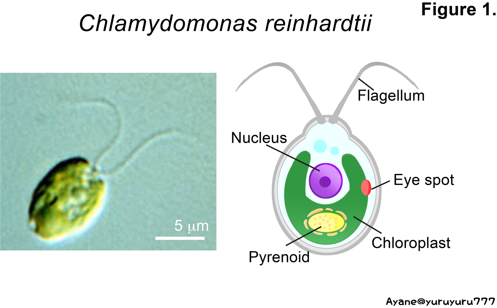
- It is easy to produce mutants because it normally grows in a haploid state.
- Classical genetic techniques such as a tetrad analysis are available.
- The databases of its genome, EST, and proteome are available, and methods for introducing exogenous genes have been established.
- Its centriole/basal body and flagellar axoneme have homologous structures to those of mammalian cells.
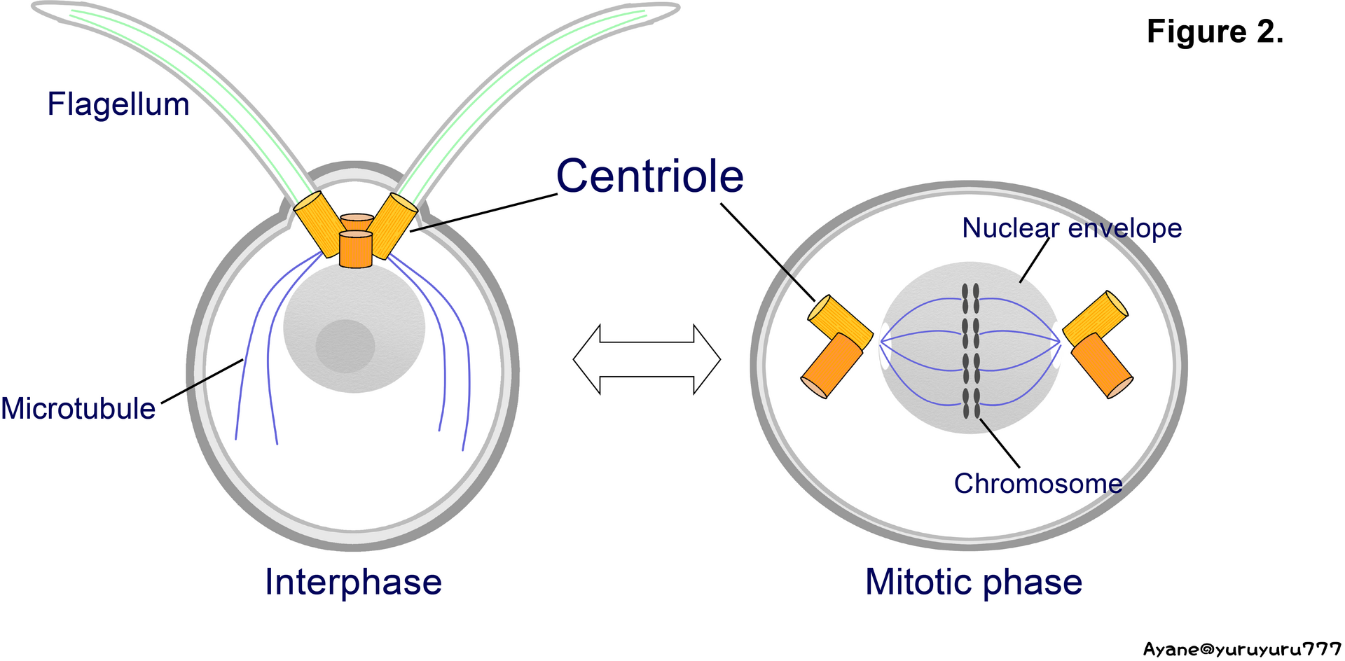
2) About centriole
Centrioles are important organelles that function as a core of the centrosome and as a base for cilia/flagellar assembly (Fig.2).
This organelle has a cylindrical structure consisting of nine triplet microtubules arranged in rotational symmetry (Fig. 3). Differently from the cytoplasmic microtubule, a dynamic singlet microtubule consisting of 13 protofilaments, the triplet is stable and contains one singlet microtubule (A-tubule) and two additional tubules (B- and C-tubules) consisting of 10 protofilaments. The A- and B-tubules of the triplet are continuous with the outer doublet microtubules of the flagellar axoneme. In other words, the centriole structure defines the 9+2 structure of the cilia/flagellar axoneme. This 9-fold symmetrical structure is highly conserved among eukaryotes; however, many questions remain about the molecular mechanisms of centriole assembly.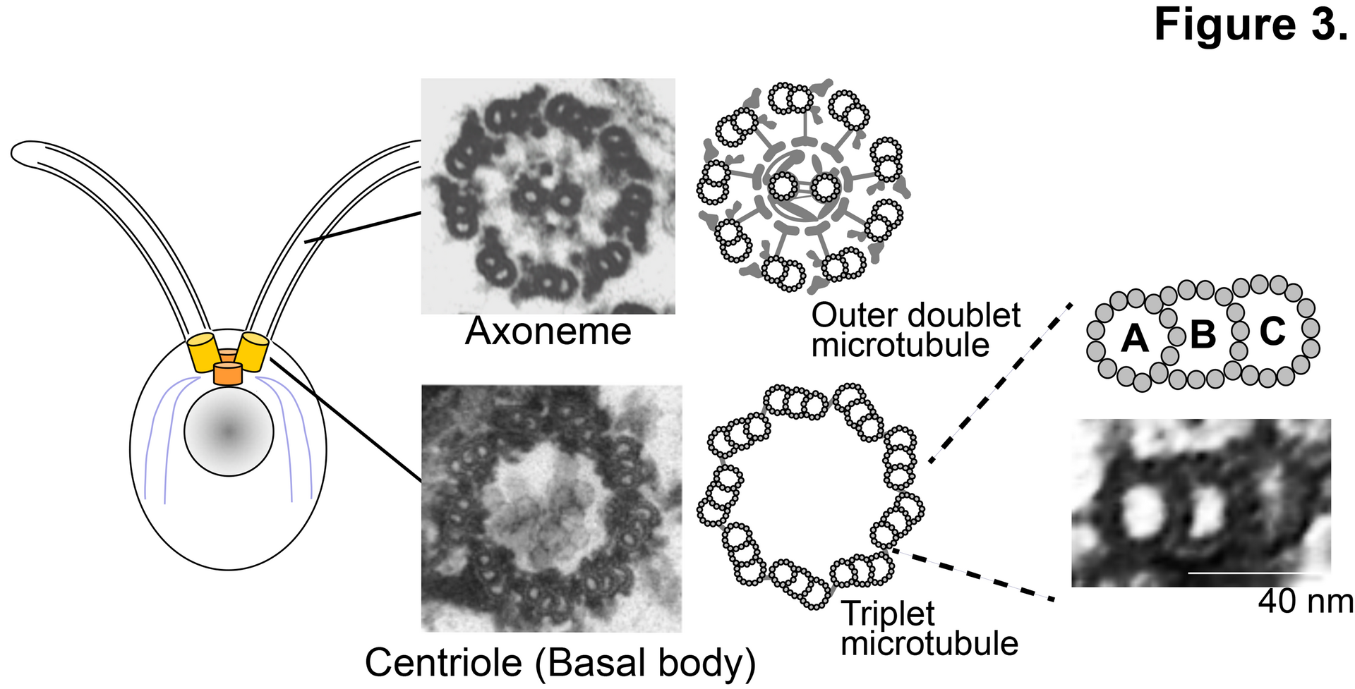
By analyzing centriole/related mutants, my research projects aim to understand the mechanisms of:
- Centriole assembly
- Triplet microtubule assembly/nucleation
- Transition zone (a region between the centriole and the flagellum) assembly and its function.
Publications
https://scholar.google.com/citations?user=wLKZTkcAAAAJ&hl=en