Invited Speakers
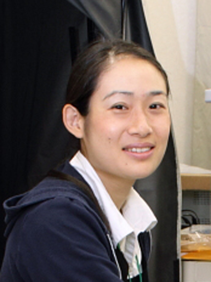
Hiroko Bannai : Laboratory of Brain Function and Structure, Nagoya University, Japan
Abstact: Approach to the brain function by imaging single molecule behavior
According to the fluid mosaic model, plasma membrane molecules such as lipids and transmembrane proteins have the ability to undergo lateral diffusion freely throughout the cell. In some cell types, however, specific membrane molecules are concentrated in cellular microdomains, by overcoming the randomizing effects of free diffusion. This polarized distribution of membrane molecules is crucial for various cell functions, thus it is important to understand the mechanism through which the cell regulates the lateral diffusion of membrane molecules. Quantum-dot single particle tracking (QD-SPT), a single molecule imaging technique using semiconductor nanocrystal quantum dots as a fluorescent probe, is a powerful tool to analyze the behavior of proteins and lipids on the plasma membrane. In this lecture, we introduce the application QD-SPT to neuroscience. We present here our recent QD-SPT experiments that allowed us to obtain further insights into the strategy and physiological relevance of membrane self-organization in neurons and astrocytes, two major component cells in the brain.
References: Bannai et al. (2006) Nature Protocols 1:2628-34; Bannai et al. (2009) Neuron 62:670-82, Niwa et al (2012) PLoS ONE 7:e36148; Arizono et al. (2012) Science Signaling 5:ra27
Biography
2013 – Present, Nagoya University, Designated lecturer,
2007 – 2013, Brain Science Institute RIKEN (Japan), JSPS research fellow, Research Scientist, Special Postdoctoral Researcher
2005 - 2007, Ecole Normale Superieure Paris (France). JSPS research fellow, Postdoctoral fellow
2000 – 2005, Brain Science Institute RIKEN (Japan), Research Scientist
1991 -2000, University of Tokyo (Japan), Ph. D (Science) 2000, M. Sci. 1997, B. Sci. 1995

Abstract: Revisiting receptor activation using multidimensional microscopy
The Epidermal Growth Factor Receptor (EGFR) is a key receptor associated with normal physiological development and functioning. When mutated or overproduced it is linked to 30% of cancers. In this pathological setting, the receptor conveys an advantage to the cancer cells probably through sending (uncontrolled) growth and survival signals. Consequently, understanding how the EGFR is activated in physiological and pathological settings is a major goal for the field and has led to a 30 year race to solve the structure of the molecule.
Together with collaborators [1,2,3,4], my team has attempted to determine the structure (assembly and conformational states) of the EGFR on cells using microscopy-based biophysical techniques. We exploit different dimensions of fluorescence imaging. Lifetime imaging microscopy in combination with Forster resonance energy transfer experiments yield insight into structural changes that occur on the 2-10 nanometre scale. Emission polarization imaging in combination with photobleaching provides insight into receptor dimerization. While fluorescence fluctuation techniques, using spatial image correlation spectroscopy, reveal the formation of receptor clusters that serve as activation “hotspots”. By combining different experimental approaches with models of reaction kinetics we can learn how receptor structure and receptor activation are correlated.
References:
1. Kozer N, Barua D, Orchard S, Nice EC, Burgess AW, Hlavacek WS, Clayton AH. Exploring higher-order EGFR oligomerisation and phosphorylation-a combined experimental and theoretical approach-Molecular Biosystems 9(7), 1849-63. (2013)
2. Kozer N, Henderson C, Jackson JT, Nice EC, Burgess AW, Clayton AHA –Evidence for extended YFP-EGFR dimers in the absence of ligand on the surface of living cells-Phys.Biol. 8: 066002 (2011)
3. Clayton, A.H.A., Walker F, Orchard SG, Henderson C, Fuchs D, Rothacker J, Nice EC, Burgess AW. Ligand- induced dimer-tetramer transition during the activation of the cell surface epidermal growth factor receptor- A multidimensional microscopy analysis-J Biol Chem. 280(34):30392-9 (2005)
4. Kozer N, Kelly MP, Orchard S, Burgess AW, Scott AM, Clayton AHA -Differential and synergistic effects of epidermal growth factor antibodies on unliganded ErbB dimers and oligomers-Biochemistry. 50(18): 3581-90 (2011)
Biography
Andrew Clayton undertook his Ph.D. studies in physical chemistry at the University of Melbourne (Prof. Ken Ghiggino) then spent time as Australian Research Council postdoctoral research fellow in the Biochemistry and Molecular Biology Department studying biophysics (Prof. Bill Sawyer). He pursued further postdoctoral studies at the Max-Planck Institute for Biophysical Chemistry in Goettingen (Dr. Thomas Jovin) as Human Frontier Science Program Long-term Fellow before returning to Melbourne initially as HFSP fellow (Prof. Tony Burgess) then National Health and Medical Research Council RD Wright Fellow and Head of the Cell Biophysics Laboratory at the Ludwig Institute for Cancer Research.
In December 2009, Dr. Clayton moved to the Centre for Microphotonics at Swinburne University of Technology where he is currently the Associate Professor of Biophotonics. His present research focuses on understanding the wiring diagram of living cells by measuring macromolecular interactions and dynamics with multidimensional microscopy. His research achievements have been recognized with several awards including Young Fluorescence Investigator from the US Biophysical Society.
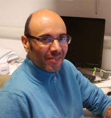
Duccio Fanelli: University of Florence, Italy
Abstract: Physics meets biology: from single molecule to population dynamics
Investigating the dynamics of individual biomolecules constitutes a topic of paramount importance of broad applied and fundamental interest. Proteins are flexible objects and exist in populations of different structures. The intrinsic ability to rearrange the spatial conformation can be instrumental in enhancing their capacity to bind other molecules and execute diverse biological functions. A quantitative theoretical understanding of proteins dynamics could be gained through classical all-atom molecular dynamics simulations. The applicability of such method is however limited in practice by computational time constraints. Alternatively, one can resort to simplified coarse grained models, the scrutinized molecule being represented as a collections of mutually interacting massive beads. Statistical mechanics provides the conceptual tools to bridge the gap between theory and experiments, making it possible to assign the physical input to a mechanical model of the protein from an ample spectrum of single molecule measurements. These approaches will be reviewed during the talk. Beyond single molecules, we will also briefly discuss the problem of modeling the dynamics of large ensemble of microscopic entities in interaction at the cellular scale. We shall in particular elaborate on the impact of molecular crowding and the role played by stochastic fluctuations.
Biography
Associate Professor in Physics at the University of Florence, Department of Physics and Astronomy. Duccio Fanelli holds a Laurea in Physics (University of Florence) and a Ph.D. in Numerical Analysis and Computer Science (KTH, Stockholm). He worked as researcher at the Karolinska Institute in Stockholm and as permanent staff member (Lecturer in Theoretical Physics) at the University fo Manchester. Fanelli has been invited professor at the University of Orleans (France) and the ENS Lyon (France). Fanelli's research interests fall in the realm of statistical mechanics and non linear physics, with applications to biology and bio-medicine. He is author of several papers in international refereed journals.
Charlotte Fournier: Okinawa Institute of Science and Technology Graduate University

Abstract
A single cell proliferates, differentiates and organises into tissues to form a multi-cellular organism. Once an organism has formed, some cells die, while others proliferate and differentiate to maintain tissue integrity and functionality. Communication between cells enables these processes to occur at the appropriate time and place. Most cells in multi-cellular organisms communicate by emitting and receiving signalling proteins, which are received and processed by a complimentary receptor protein located in the cell membrane. The epidermal growth factor receptor is an example of such a cell membrane receptor. During development the epidermal growth factor receptor controls epithelial cell proliferation and differentiation within several organs; the skin, lungs, liver and gastrointestinal tract. In fully developed organisms, for example humans, dysregulation of the epidermal growth factor receptor is often found in carcinomas - cancers of epithelial cell origin. We are interested in the dynamic arrangement and mobility of the epidermal growth factor receptor within the cell membrane using total internal reflection fluorescence microscopy and single molecule detection and tracking algorithms.
Biography
Charlotte Fournier studied Medicine at Cardiff University, before working in General Internal Medicine at the University Hospital of Wales and the Royal Devon and Exeter Hospital in England. She then moved to the University of Bristol, United Kingdom, as a Medical Demonstrator, teaching Medical Physiology to medical, dental and veterinary science students. Following this she started a D.Phil in Biophysics at the University of Oxford under Professor Mark Leake. She is now a research scientist at the Okinawa Institute for Science and Technology.

Abstract: Quantitative single-molecule super-resolution microscopy of cellular structures
In the fluorescence microscopy field, much interest has focused on new super-resolution techniques (collectively known as PALM/STORM, STED, SIM and others) [1] that demonstrated to bypass the diffraction limit and provide a spatial resolution reaching a near-molecular level. With these techniques, it has become possible to image cellular structures in far greater detail than ever before. Single-molecule based methods such as photoactivation-localization microscopy (PALM) [1], stochastic optical reconstruction microscopy (STORM) [2] and directSTORM (dSTORM) [3] employ photoswitchable fluorophores and single-molecule localization to generate a super-resolution image.
The important next step is to obtain reliable quantitative information from super-resolution images of cellular structures. Single-molecule super-resolution techniques can be applied to study the aggregation of membrane proteins and the nano-structural organization of HIV-1 envelope protein in intact cells [4] and single viruses. Furthermore, single-molecule counting of nucleosome-associated proteins [5] and transcription-associated proteins [6] will be presented.
[1] E. Betzig et al., Science 2006, 313, 1642-1645.
[2] M. J. Rust et al., Nature Methods 2006, 10, 793-795.
[3] M. Heilemann et al., Angewandte Chemie 2008, 47, 6172-6176.
[4] W. Muranyi et al., Plos Pathogens 2013, 9(2), e1003198
[5] Lando et al., Open Biology 2012, 2:120078.
[6] Endesfelder et al., Biophys J 2013, 105, 172-181.
[6] Endesfelder et al., Biophys J 2013, 105, 172-181.
Research interest
The main focus of my research is to develop advanced microscopic and spectroscopic techniques, such as single-molecule fluorescence and super-resolution microscopy techniques, and to apply these to study cellular structures with high spatial and temporal resolution. Specifically, I am interested in the structural organization of proteins and biomolecules at small spatial scales, including membrane proteins, nucleic acids and machines of transcription and replication.
Biography
Mike Heilemann holds a diploma in Chemistry from the University of Heidelberg. He finished his PhD under the supervision of Prof. Markus Sauer in de Department of Physics at the University of Bielefeld in 2005. From 2005 to 2007, he worked as a postdoctoral research fellow in the Physics Department at the University of Oxford, in the Section of Bionanotechnology. In 2008, he was awarded junior research group funding by the BMBF FORSYS program. In 2011, he was appointed assistant professor (W2) at the University of Würzburg. In 2012, he was appointed full professor (W3) at the University of Frankfurt, where he is now heading the department of Single Molecule Biophyiscs.
Akihiro Kusumi : Institute for Frontier Medical Sciences, Kyoto University, Japan

Abstract: Organizing principles of the plasma membrane for signal transduction: Three-tiered hierarchical meso-scale domain architecture revealed by single-molecule tracking
Single-molecule imaging and tracking techniques that are applicable to living cells are revolutionizing our understanding of the plasma membrane dynamics, structure, and signal transduction functions. The plasma membrane is considered the quasi-2D non-ideal fluid that is associated with the actin-based membrane-skeleton meshwork, and its functions are likely made possible by the mechanisms based on such a unique dynamic structure, which I call membrane mechanisms.
My group is largely responsible for advancing high-speed single-molecule tracking of membrane-associated molecules. Based on the observations made by this approach, I propose that the cooperative action of the hierarchical three-tiered meso-scale (2-300 nm) domains, (1) actin-membrane-skeleton induced compartments (40-300 nm), (2) raft domains (2-20 nm), and (3) dynamic protein complex domains (3-10 nm), is critical for the membrane function. In particular, the actin-based membrane skeleton distinguishes the plasma membrane from the Singer-Nicolson-type membrane.
Education:
1971/4-1975/3 Undergraduate: Major field, Biophysics
Dept. of Biophysics, Faculty of Science, Kyoto University
1975/3 B. Sc. in Biophysics
1975/4-1980/3 Graduate: Major field, Biophysics
Dept. of Biophysics, Faculty of Science, Kyoto University
1980/3 D. Sc. in Biophysics
Work Experience:
1980/4-1982/10 Research Associate (National Biomedical ESR Center, Medical College of Wisconsin)
1982/11-1984/4 Research Fellow (Department of Biology, Princeton University)
1984/5-1988/4 Assistant Professor (Department of Biophysics, Faculty of Science, Kyoto University)
1984/5-1994/8 Visiting Professor, Department of Radiology and Biophysics (Medical College of Wisconsin)
1988/5-1997/4 Associate Professor (Graduate School of Arts and Sciences, The University of Tokyo)
1997/5-2004/12 Professor (Department of Biological Science, Nagoya University)
1998/10-2005/3 Project Leader (Membrane Organizer Project, ERATO/SORST-JST)
2005/4-2010/3 Project Leader (Membrane Mechanisms Project, ICORP-JST)
2005/1-Present Professor (Institute for Frontier Medical Sciences, Kyoto University)
2007/10-Present Professor (Institute for Integrated Cell-Material Sciences (iCeMS)), Kyoto University
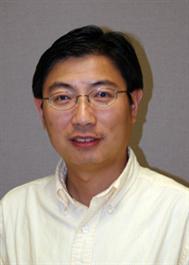
Jun Liu : University of Texas Medical School, USA
Abstract:
Molecular machines are large macromolecular assemblies that underlie the fundamental biological processes in cells. My laboratory is interested in three-dimensional structure and function of molecular machines in living cells. We developed a unique high-throughput cryo-electron tomography (cryo-ET) system to visualize those biologically important molecular machines in their native cellular environment [1-6]. In order to achieve high resolution in situ structures, we specifically choose Lyme disease spirochete (Borrelia Burgdorferi) [3, 6] and very tiny E. coli minicell [4, 5] as two main model systems. In this talk, I will present our recent progress in understanding flagellar assembly, bacterial motility, and bacteriophage infection. Recent developments and potentials in cryo-ET will be discussed in the context of its exciting applications.
[1] Liu J, Taylor DW, Krementsova EB, Trybus KM, Taylor KA: Three-dimensional structure of the myosin V inhibited state by cryoelectron tomography, Nature 442:208-211, 2006.<http://www.ncbi.nlm.nih.gov/pubmed/16625208>
[2] Liu J, Bartesaghi A, Borgnia M, Sapiro G, Subramaniam S: <http://www.ncbi.nlm.nih.gov/pubmed/18668044> Molecular architecture of native HIV-1 gp120 trimers<http://www.ncbi.nlm.nih.gov/pubmed/18668044>, Nature 455:109-113, 2008.<http://www.ncbi.nlm.nih.gov/pubmed/18668044>
[3] Liu J, Lin T, Botkin DJ, McCrum E, Winkler H, Norris SJ: Intact Flagellar Motor of Borrelia burgdorferi Revealed by Cryo-Electron Tomography: Evidence for Stator Ring Curvature and Rotor/C Ring Assembly Flexion, J Bacteriol 191(16):5026-36, 2009.<http://www.ncbi.nlm.nih.gov/pubmed/19429612>
[4] Liu J, Hu B, Morado DR, Jani S, Manson MD, Margolin W: Molecular architecture of chemoreceptor arrays revealed by cryoelectron tomography of Escherichia coli minicells, Proc Natl Acad Sci U S A 109(23):E1481-8, 2012.<http://www.ncbi.nlm.nih.gov/pubmed/22556268>
[5] Hu B, Margolin W, Molineux IJ, Liu J: The Bacteriophage T7 Virion Undergoes Extensive Structural Remodeling during infection, Science 339(6119):576-9, 2013.<http://www.ncbi.nlm.nih.gov/pubmed/23306440>
[6] Zhao X, Zhang K, Boquoi T, Hu B, Motaleb MA, Miller K, James M, Charon NW, Manson MD, Norris SJ, Li C, Liu J: Cryo-Electron Tomography Reveals the Sequential Assembly of Bacterial Flagella in Borrelia burgdorferi, Proc Natl Acad Sci U S A, 110(35): 14390-5.<http://www.ncbi.nlm.nih.gov/pubmed/?term=23940315>
Biography:
Assistant Professor at the University of Texas-Houston Medical School, Houston, Texas. He got his Ph.D in Physics and Electron Microscopy at Institute of Physics, Chinese Academy of Sciences in Beijing. He then started learning Electron Tomography with Prof. Ken Taylor at Florida State University. As a staff scientist at National Institutes of Health (NIH), he worked on the molecular architecture of HIV envelope spike. Now he is a Principle Investigator in several projects supported by NIH and Welch Foundation.
Tomoko Masaike : Tokyo University of Science, Japan

Abstract: Imaging motions, functions, and reactions of single molecules
The aim of our research is to investigate how motions and chemistry of single protein molecules or organelles are coupled with physiological functions.
First, dynamic motions of a motor protein F1-ATPase and an organelle airway epithelial cilium were detected through beads under the optical microscope. Trajectories of these probes were analyzed to quantify rotational or beating motions.
Secondly, local conformational changes of proteins were monitored by detecting orientation of fluorophores attached to secondary structures of proteins under TIRF microscopy with polarization modulation. We are trying to explore conformations of ATPase enzymes F1-ATPase and Ca2+-ATPase. The optical system that enables determining orientations of fluorescent probes in 3D is under development. Mapping local conformational changes of multiple regions will clarify how subtle conformational changes at the catalytic sites are propagated to distant domains.
Finally, chemistry of single molecules is visualized. Our approach is to detect release of inorganic phosphate from enzymes by fluorescence increase under the microscope. Rhodamine-labeled phosphate binding protein encapsulated in microchambers increases fluorescence intensity as it binds inorganic phosphate, a product of ATP hydrolysis.
These observation systems will be integrated and contribute to understanding of biological systems. Our future direction is to apply these techniques to in vivo imaging.
Biography
Tomoko Masaike, Ph. D. is a lecturer in the Department of Applied Biological Science, Tokyo University of Science from 2013 and concurrently, a researcher of JST’s PRESTO project, Structural Life Science. Her research areas are Biophysics and Biochemistry. She is especially interested in the interplay between chemical reactions, conformational changes, and functions of biological molecules. In order to study them, she tries to link single-molecule observation under the optical microscope to ensemble measurements of numerous molecules.
She initiated her studies on conformational changes of a rotary molecular motor FoF1-ATP synthase using a fluorometer when she became a master’s course student in Prof. Masasuke Yoshida’s group in Tokyo Institute of Technology in 1996, where she later earned her Ph. D. in 2002. Then she joined ERATO Yoshida ATP system project, JST as a postdoctoral fellow and started collaboration with Dr. Nishizaka for single-molecule observation of local conformational changes of F1-ATPase under the microscope. In 2005, she joined Prof. Nishizaka’s group in Gakushuin University as a research associate and continued collaboration. In 2008, local conformational changes of this enzyme were correlated with rotation of the central shaft by simultaneous observation under TIRF microscopy with polarization modulation
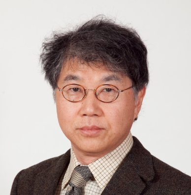
Shigetoshi Oiki : Department of Medicine, Fukui University, Japan
Abstract: Spatio-Temporal Interplay of the Single KcsA Potassium Channel in the Membrane.
For the ion channel, the lipid membrane is both the supporting scaffold and the physicochemical matrix, where channels diffuse more or less freely and interplay with lipid molecules. Here we applied various single-molecule measurement techniques and examined the pH-dependent KcsA channel from the level of sub-molecular conformational changes to supra-molecular clustering–dispersion dynamics in the membrane. Upon gating, the KcsA channel undergoes twisting conformational changes around the symmetry axis of the channel (Shimizu et al., Cell 2008), which is accompanied with the substantial shortening and elongation of the channel along the longitudinal axis (Sumino et al. Sci. Rep. 2013). The gating is strongly affected by membrane lipids, and the KcsA channel senses compositional changes through the lipid-sensing antenna (Iwamoto & Oiki, PNAS 2013). Thus, these global conformational changes upon gating should lead to different mechanical interactions with the surrounding membrane. We found that in the open conformation, channels diffused freely on the lipid bilayer as singly isolated channels, but in the closed conformation they were self-assembled. Reversible clustering–dispersion dynamics was captured (Sumino et al. JPCLett. 2014). The spatio–temporal interplay of channel proteins on the membrane evolve, and dynamic pictures of the functioning channel are now integrating.
Biography
2003-present: Professor, Department of Molecular Physiology and Biophysics, Faculty of Medical Sciences, University of Fukui
1998-2003: Professor, Department of Physiology, Department of Physiology, Fukui Medical University
1992-1998: Associate Professor, Department of Cellular and Molecular Physiology, National Institute for Physiological Sciences
1989-1992: Assistant Professor, Department of Physiology, Kyoto University Faculty of Medicine
1988-1989: Research associate, Department of Physiology and Biophysics, Cornell University Medical College (Mentor: Olaf S. Andersen)
1986-1988: Postdoctoral fellow, Department of Neurosciences, Roche Institute for Molecular Biology (Mentor: Mauricio Montal and Richard Horn)
1986-1986: Research Associate, Department of Physiology, Kyoto University Faculty of Medicine
1978-1982: Resident, Department of Anesthesiology, Kyoto University Hospital
Education
1982-1986 Kyoto University Faculty of Medicine, Ph.D. in Physiology
1972-1978 Kansai Medical University, M.D.
Social activities
2008-present: Editorial board, the e-Journal “BIOPHYSICS”
2014-present: Executive board member, Physiological Society of Japan
Awards
2012: Prizes for Science and Technology, Research Category. The Commendation for Science and Technology by the Minister of Education, Culture, Sports, Science and Technology
Abstract
Behaviors of cells are regulated by protein reactions inside cells. The numbers of molecules involved in unitary reaction steps in the reaction networks are small, and quantitative information of these unitary reaction steps are largely lacking. Therefore, single-molecule measurement is useful and may be indispensable technology for studying cell signaling reactions quantitatively to understand cell behaviors. Using single-molecule technologies both in vitro and in living cells, we are studying cell fate decision carried out by ErbB-RAS-MAPK system and PAR system, both of which are complicated protein reaction networks inside cells. ErbB-RAS-MAPK system is regulating cell proliferation, differentiation, death, and carcinogenesis. PAR system is responsible for establishment and maintain of cell polarization. In this talk, I would like to introduce the technology and usage of single-molecule measurements of intracellular reaction networks, and show complex properties of protein reactions observed using single-molecule technologies. Combination of quantitative single-molecule analysis and mathematical modeling is revealing how cell dynamics is produced from protein reaction systems.
Biography
Yasushi Sako obtained his PhD in 1990 from Kyoto University. After worked at Tokyo University (1989-1996), Nagoya University (1997), and Osaka University (1997-2005) as assistant or associate professor, he moved to RIKEN (Institute of Physics and Chemistry) in 2006 to start Cellular Informatics Laboratory as the chief scientist. From 2010, he is a guest professor of Graduate School of Frontier Biosciences, Osaka University, concurrently with RIKEN. His research interests focus on intracellular reaction networks of biosignaling, structure and function of proteins, and optical microscopy.
Takayuki Uchihashi : Kanazawa University, Japan
Abstract: Visualization of Single Molecule Dynamics at Work with High-Speed Atomic Force Microscopy
Biological molecules fulfil a wide variety of unique functions. Their functions are essentially elicited from conformational change and/or interactions with other molecules which are often triggered by binding of ligand/substrate and changes in the external environment. Therefore, studying dynamic processes of individual molecules, for example, how molecules undergo conformational change and how molecules interact, is indispensable to understand the dynamic relationship between structure and function in biological molecules. Nevertheless, a tool with an ability to directly detect and track conformational change in real time has not been available.
Atomic force microscopy (AFM) is a versatile technique to study nanoscale structures of materials under various environments. One of the most coveted new functions of AFM is “fast recording” because it allows the observation of dynamic processes occurring at the nanoscale. The visualization of dynamic processes provides direct and deep insights into the target objects and phenomena under the microscope. This new capability of observation should open a new opportunity to reveal essential mechanisms of working proteins.
Biography
Takayuki Uchihashi received his B. Sc., M.Sc. in Physics from Hiroshima University, Japan in 1993 and 1995, respectively. In 1998, he obtained Dc. Eng. in Electronics from Osaka University, Japan. From 1998 to 2000, he worked at Joint Research Center for Atom Technology (JRCAT) in Tsukuba as a research associate. In 2000, he joined the Department of Electronic Engineering, Himeji Institute of Technology. In 2002, he moved to Trinity College in Dublin and worked as a senior researcher. In 2004, he joined the Physics Department, Kanazawa University, and since 2006 he is an associate professor. His research interests include the instrumentation of scanning probe microscopy and its application to biological science.
Vladana Vukojevic : Karolinska Institutet, Sweden
Abstract
Living organisms control the concentration, spatial distribution and temporal behavior of bioactive molecules through molecular interactions and transporting processes. In order to understand how local molecular interactions are integrated to form specific biochemical pathways and control their cellular dynamics, kinetics of molecular interactions needs to be quantitatively characterized in live cells. In the talk entitled “Quantitative Live Cell Biochemistry. Dynamic regulation of biological processes through dimerization” Dr. Vukojević will introduce Fluorescence Correlation Spectroscopy (FCS), a quantitative method with single-molecule sensitivity and give examples of its application for the study of kinetic regulatory mechanisms in live cells. Topics that will be discussed build further on published studies of the dynamic lateral organization of G Protein-Coupled Receptors (GPCRs) [1,2,3] and gene transcription regulation by transcription factors [4,5].
1. Tobin SJ, Cacao EE, Hong DWW, Terenius L, Vukojević V, Jovanović-Talisman T. Nanoscale effects of ethanol and naltrexone on protein organization in the plasma membrane studied by Photoactivated Localization Microscopy (PALM). PLoS One, 2014 Feb 4
2. Gregorsson Lundius E, Vukojević V, Hertz E, Stroth N, Cederlund A, Hiraiwa M, Terenius L, Svenningsson P. GPR37 Trafficking to the Plasma Membrane Regulated by Prosaposin and GM1 Gangliosides Promotes Cell Viability. J. Biol. Chem. 2013 in press (doi: 10.1111/jnc.12081. Epub 2012 Dec 6.)
3. Vukojević V, Gräslund A, Bakalkin G. Fluorescence imaging with single-molecule sensitivity and fluorescence correlation spectroscopy of cell-penetrating neuropeptides. Methods. Mol. Biol. 2011 789:147-170.
4. Vukojević V, Papadopoulos DK, Terenius L, Gehring W, Rigler R, Quantitative study of synthetic Hox transcription factor–DNA interactions in live cells, Proc Natl Acad Sci USA 2010, 107:4087-4092.
5. Papadopoulos DK, Vukojević V, Adachi J, Terenius L, Rigler R, Gehring W. Function and specificity of synthetic Hox transcription factors in vivo. Proc Natl Acad Sci USA 2010, 107:4093-4098.
Biography
Dr. Vladana Vukojević, born in Belgrade, Serbia, has acquired B.Sc., M.Sc. and Ph.D. degrees in Physical Chemistry at the Faculty of Physical Chemistry, University of Belgrade, Serbia. She commenced her research career at the H. C. Ørsted Institute, Copenhagen, Denmark, in 1990, and her academic career began in 1992 at the Faculty of Physical Chemistry, University of Belgrade, Serbia, where she was Associate Professor of Biophysical Chemistry until 2005. She has joined Karolinska Institute, Stockholm, Sweden in 2003, where she is Associate Professor of Biochemistry since 2011. Her research focuses on self-organization of biochemical systems and the understanding of kinetic mechanisms through which local molecular interactions are integrated to build specific biochemical pathways. She uses fluorescence microscopy imaging and correlation spectroscopy to identify and quantitatively characterize in live cells self-regulatory kinetic mechanisms underlying vital biological functions such as gene transcription and cellular signaling. Dr. Vukojević is coauthor of 50 papers published in peer reviewed journals.
Paul WISEMAN : Departments of Chemistry & Physics McGill University, Canada

Abstract: Bridging the gap between ensemble and single molecule fluorescence methods with image correlation spectroscopy
Image correlation methods are an extension of fluorescence fluctuation spectroscopy that can measure protein-protein interactions and macromolecular transport properties from input fluorescence microscopy images of living cells. These approaches are based on space and time correlation analysis of fluctuations in fluorescence intensity in time series images recorded on a laser scanning or total internal reflection fluorescence (TIRF) microscope. Spatio-temporal image correlation spectroscopy (STICS) measures vectors of protein flux in cells based on the calculation of a spatial correlation function as a function of time from an image time series. We will describe the application of STICS and its two color extension, spatio-temporal image cross-correlation spectroscopy (STICCS), for measuring transport maps of adhesion related macromolecules integrin, alpha-actinin, paxillin, talin, and vinculin within, or associated with the basal membrane in living U2OS and CHO cells as well as during dynamic turnover of podosomes in dendritic immune cells. We will also highlight recent results from a new form of reciprocal (k-) space ICS (kICS) and compare single molecule tracking and kICS measurements of transport signaling receptors in the cell membrane.
Biography
Paul Wiseman obtained his B.Sc. (Honours) from St. Francis Xavier University in 1989 and Ph.D. in Chemistry from the University of Western Ontario in 1995 where he held a NSERC postgraduate research fellowship. Afterwards he was awarded a Japan Society for the Promotion of Science postdoctoral fellowship at the University of Tokyo and Nagoya University (with Prof. Akihiro Kusumi), and later became a LJIS interdisciplinary postdoctoral research fellow at the University of California, San Diego (with Prof. Kent Wilson). In 2001, Paul started as an assistant professor jointly appointed in the Departments of Chemistry and Physics at McGill University, was promoted to associate professor in 2007, and to full professor in 2013. His research focuses on biophysics measurements of protein interactions and transport in living cells and neurons. Wiseman has published a total of 59 peer reviewed publications since starting at McGill University in 2001 including 2 journal cover articles and 6 Faculty of 1000 Biology Selections. He was awarded the Young Fluorescence Investigator award in 2005 by the Biophysical Society and the 2009 Keith Laidler Award in Physical Chemistry by the CSC. In 2007 he was awarded the Yaffe Teaching Award and the Principal’s Prize for Teaching by McGill as well as the J.D. Jackson Teaching Award in Physics in 2012. In 2012 Wiseman was named the Otto Maass Chair in Chemistry at McGill University.
Masataka Kinjo : Hokkaido University, Japan

Abstract: Study of dimer formation of transcription factor using fluorescence cross-correlation spectroscopy in the living cell
Two-laser-beam fluorescence cross-correlation spectroscopy (FCCS) is promising technique that provides quantitative information about the interactions of biomolecules. Using FCCS, we determined the dissociation constant (Kd) of the p50/p65 heterodimer, homodimer and also homodimer of glucocorticoid receptor (GR) in living cells. GR is a well-known ligand-dependent transcriptional regulatory protein. The classical view is that unliganded GR resides in the cytoplasm, translocates to the nucleus upon ligand binding, and then associates with the GRE to transactivate a specific gene. It is still a puzzle whether GR forms a dimer in the cytoplasm or nucleus before or after DNA binding in the nucleus. Our result suggested that high values of Kd in nucleus and also cytoplasmic dimerization. These findings support the existence of a dynamic monomer pathway and regulation of GR in both the cytoplasm and nucleus.
Biography
Masataka Kinjo obtained his PhD in 1985 from Jichi Medical School, Japan. After worked at Research Institute of Applied Electricity, Hokkaido University as assistant and associate professor, he became professor at Faculty of Advanced Life Science, Hokkaido University on 2007. He spent two years in Karolinska Institute as a guest researcher from 1991 to 1993.
His main research field is biophysics and cell biology by using several kinds of advanced fluorescence microscopy including fluorescence correlation spectroscopy (FCS), fluorescence cross correlation spectroscopy (FCCS) and multi-point total internal reflection FCS (MP-TIR-FCS).
Member of Review Committee for HFSP Research Grants 2011-now.
Thorsten Wohland : National University of Singapore

Abstract
Imaging FCS can measure millions of points simultaneously with millisecond time resolution, or thousands of points with a time resolution down to the microsecond range. The measurements of an FCS image is sufficiently fast (< 20 s) to allow the recording of FCS time series, observing the dynamics of cells and bilayers over an extended period of time. In this seminar we will discuss the theoretical and practical aspects of imaging FCS and its implementation using total internal reflection (TIR) or single plane illumination microscopy (SPIM). We used imaging TIR-FCS (ITIR-FCS) to observe the action of monomeric human islet polypeptide (hIAPP) on live cell membranes over a time span of one hour using FCS time lapse movies. SPIM-FCS provides similar data to ITIR-FCS but can perform measurements in true 3D samples. We applied SPIM-FCS to a range of different proteins (fluorescent proteins, transcription factors and Rho-GTPases) in live cells and in zebrafish embryos to determine location dependent diffusive behavior. Imaging FCS provides sufficient time resolution to observe molecular processes in cells and organisms similar to confocal FCS. But its inherent multiplexing allows collecting many more data points simultaneously with reduced optical damage, allowing more measurements per sample.
Biography
Thorsten Wohland studied Physics at the Technical University of Darmstadt and the University of Heidelberg in Germany from 1989-1995. He completed his diploma thesis in physics at the European Molecular Biology Laboratory (EMBL) in Heidelberg, Germany, under the supervision of Ernst H.K. Stelzer. In 1997 he joined the group of Prof. Horst Vogel at the Swiss Federal Institute of Technology in Lausanne (EPFL), Switzerland. In 2000 he obtained his doctoral degree for the study of theoretical and practical aspects of fluorescence correlation spectroscopy (FCS). Following two years in the group of Richard N. Zare at Stanford in the USA working on single molecule detection and protein immobilization he started as Assistant Professor at the National University of Singapore (NUS) in June 2002. At NUS he developed several new fluorescence correlation spectroscopy methods, imaging total internal reflection FCS (ITIR-FCS), single plane illumination microscopy FCS (SPIM-FCS), and single wavelength fluorescence cross-corrleation spectroscopy (SW-FCCS), which allowed for the first time to take correlation images of live cells and quantitatively measure affinity constants of bimolecular interactions in live organisms, respectively. His current research aims at the application of these methods to biological problems and at the combination of super-resolution microscopy with imaging FCS.


