FY2019 Annual Report
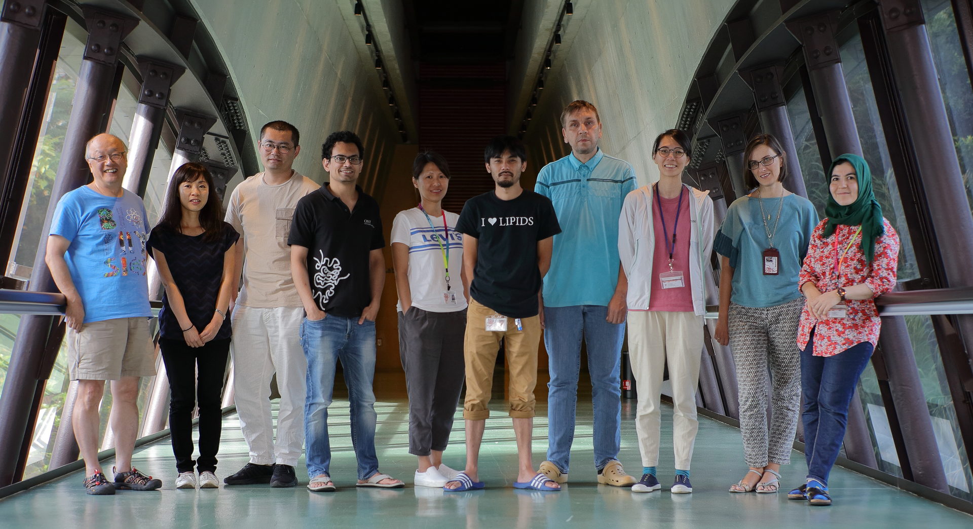
Membrane Cooperativity Unit
Professor Akihiro KUSUMI

Abstract
The Membrane Cooperativity Unit is dedicated to (1) methodology development for single-molecule imaging and manipulation at nanometer precisions in living cells, with a special attention paid to high time resolutions (ultrafast single fluorescent-molecule tracking), as well as (2) revealing meso~nano-scale processes in signal transduction in/on the cell membrane and the formation and remodeling of the neuronal network, by using the developed single-molecule techniques. The smooth liaison between physics/engineering and biomedicine is a key for our research.
We recently developed a new ultrafast (indeed the world-fastest) camera system for single fluorescent-molecule imaging. This system allows us to image single fluorescent molecules at time resolutions down to 20 µs (1,650x faster than normal video rate; many single molecules at the same time in the view-field), which represents the ultimate speed using the presently-available fluorescent molecules. At the same time, it allows us to simultaneously observe two or three molecular species. We also developed a method for observing single fluorescent molecules as long as 12,000 frames (~7 min at video rate, as compared to the general ~10 s, i.e., world longest).
The plasma membrane is the outermost cellular membrane, which encloses the entire cell. It is critically important for the cell - the fundamental unit of life - because it defines the space for it. The plasma membrane exchanges information, energy, and substances with the outside world, and we pay special attention to the mechanism for signal transfer from outside to inside the cell, a function generally called “signal transduction”. In the signal transduction process, the plasma membrane works like a sensor + computer + effector.
The Membrane Cooperativity Unit strives to understand how the plasma membrane works at very fundamental levels, based on unique insights we obtain by applying single-molecule tracking methods to nano~meso-scale processes occurring in signal transduction and neuronal network formation/modulation in the cellular plasma membrane. More specifically, we are now revealing the mechanisms by which the metastable molecular complexes and meso-scale membrane domains, including membrane compartments, raft domains, and protein oligomers, form and work in concert to enable signal transduction and synapse formation/modulation in/on the plasma membrane.
Based on the unique insight and understanding we obtain for these important cellular processes, we hope to develop new types of systems molecular biology.
1. Staff
- Dr. Amine Betul Nuriseria Aladag, Post Doctoral Scholar
- Dr. HooiCheng Lim, Post Doctoral Scholar
- Dr. Taka-Aki Tsunoyama, Post Doctoral Scholar
- Dr. Peng Zhou, Post Doctoral Scholar
- Dr. Irina Meshcheryakova, Technician
- Ms. Limin Chen, Technician
- Ms. Hiroko Hijikata, Technician
- Ms. Aya Nakamura, Technician
- Ms. Yuri Nemoto, Technician
- Mr. Alexey Yudin, Technician
- Mr. Yoshifumi Maeda, Research Assistant (Part-time)
- Mr. Shogo Miyagi, Research Assistant (Part-time)
- Mr. Masaya Negawa, Research Assistant (Part-time)
- Mr. Tatsuhiro Nishi, Research Assistant (Part-time)
- Mr. Takaya Shimabukuro, Research Assistant (Part-time)
- Mr. Hiroto Uchihara, Research Assistant (Part-time)
- Ms. Sachie Matsuoka, Research Unit Administrator (also for Laurino Unit)
- Ms. Miwako Tokuda, Research Unit Administrator
- Dr. Akihiro Kusumi, Professor
2. Collaborations
2.1 Revealing the dynamics, structure, and function of metastable signaling molecular complexes by single-molecule imaging
- Description: Developing ultrafast 3D single-molecule imaging, and applying it to revealing the dynamics and formation mechanism of the signaling complex in synaptic signaling, Fcepsilon signaling, focal adhesion architecture and signaling, and GPI-anchored proteins’ raft-based signaling
- Type of collaboration: Joint research
- Researchers:
- Dr. Takahiro Fujiwara, Associate Professor, Institute for Integrated Cell-Material Sciences (iCeMS), Institute of Advanced Studies, Kyoto University
- Dr. Kenichi Suzuki, Professor, G-CHAIN, Gifu University
2.2 Unraveling the large-scale molecular-species selective diffusion barriers in the axonal initial segment in the neuron using ultrafast single-molecule imaging
- Description: By applying ultrafast single-molecule imaging and ultrafast single-molecule localization microscopy developed by us, we try to unravel the large-scale molecular-species selective diffusion barriers in the axonal initial segment in the neuron
- Type of collaboration: Joint research
- Researchers:
- Dr. Takahiro Fujiwara, Associate Professor, Institute for Integrated Cell-Material Sciences (iCeMS), Institute of Advanced Studies, Kyoto University
2.3 Elucidation of dynamics and formation mechanisms of cellular signaling complexes by developing new single particle tracking methods
- Description: Developing fluorescent probes for their applications to single-molecule imaging in living cells, and by using the developed probes, elucidating dynamics and formation mechanisms of cellular signaling complexes induced by various intercellular signaling molecules and alien antigens, including (non-pathogenic) viruses
- Type of collaboration: Joint research
- Researchers:
- Dr. Dai-Wen Pang, Professor
- Mr. Bo Tang, PhD candidate
- Ms. Dan-dan Fu, PhD candidate
- Ms. Jing Li, PhD candidate
College of Chemistry and Molecular Sciences, Wuhan University, P. R. China
2.4 Revealing the raft-based signaling by using single-molecule imaging
- Description: Revealing the signal transduction mechanisms of GPI-anchored proteins and raft-based signaling by using single-molecule imaging
- Type of collaboration: Joint research
- Researchers:
- Dr. Ikuko Koyama-Honda, Lecturer, Graduate School of Medicine, The University of Tokyo
2.5 Elucidating the functions of plasma membrane compartmentalization
- Description: Elucidating how the signal transduction functions of the plasma membrane is regulated using the actin-based compartmentalization of the plasma membrane, using ultrafast single-molecule imaging-tracking and super-resolution microscopy
- Type of collaboration: Joint research
- Researchers:
- Dr. Pakorn Tony Kanchanawong, Professor, Mechanobiology Institute, The National University of Singapore
3. Activities and Findings
3.1 Revealing metastable signaling molecular complexes by developing new single-molecule imaging methods
In this project, we develop new single fluorescent-molecule imaging-tracking methods and new fluorescent probes, including ultrafast (world’s fastest) single-molecule imaging, methods for suppressing photobleaching and photoblinking for very long single molecule tracking (several minutes) in living cells, and new fluorescent lipid probes that behave very much like their parent endogenous lipid molecules. By applying the developed methods and probes, we try to reveal the dynamics and formation mechanism of metastable signaling molecular complexes in the context of synaptic signaling, Fcepsilon signaling, focal adhesion architecture and signaling, GPI-anchored proteins’ raft-based signaling, and ganglioside functions in the formation and function of raft domains.
3.2 Unraveling the regulation mechanisms for the synaptic structural plasticity by observing dynamics, assembly, and function of neuronal receptors using super-resolution single-molecule imaging and tracking
In this project, we try to understand the mechanisms by which structural synaptic plasticity is induced. To accomplish this goal, we examine, at the level of single molecules, molecular interactions and dynamics in hippocampal neurons in culture. In particular, we examine the dynamic equilibrium of monomers, dimers, oligomers, and clusters of neurotramsmitter receptors and other neuronal molecules, the molecules’ cooperative interactions, including the possibility of phase separation, as well as their entrance-exiting dynamics into and out of the synapses.
In 2019, we have found that AMPA receptors form and disintegrate continually, within a fraction of a second, rather than being a very stable entity, as previously thought. These findings, published in Nature Communications (Morise et al., 2019), may help clarify early stages of synaptic plasticity: how the brain changes the neuronal circuits to learn and memorize. The research may also have pharmacological applications in the treatment of epilepsy. The following is the brief explanations of this research result.
Nerve cells or neurons communicate with one another in their specialized surface domains called synapses. The key event for learning and memory is the modulation of this communication in the synapse, called synaptic plasticity. In the synapse, a neuron emits chemical messengers called neurotransmitters, and the apposed neuron receives them using tiny structures called receptors. The AMPA receptor localized in the synapse plays a critical role in synaptic plasticity. However, how AMPA receptors were put to work has yet to be fully understood.
AMPA receptors are composed of four molecules or subunits, which unite to form structures called tetramers. There are four kinds of subunits, GluA1, 2, 3, and 4, to form tetramers, and therefore, there could be 256 kinds of AMPA receptors. It has been widely believed that these tetramers originate in the endoplasmic reticulum, which is often referred to as the cell’s manufacturing center, and then migrate to the synapses, and that once the tetramers are produced, they are stable for hours and days.
However, if this were true, it would create a large problem for neurons. The synapse needs AMPA receptor tetramers with different combinations of subunits as the brain develop and its neuronal circuits change by new information, and this occurs all the time. If AMPA receptor tetramers are stable, when the synapse requires different kinds of AMPA receptor tetramers, they have to be newly synthesized from scratch and brought to the synapse or the neuron has to keep large stocks of great varieties of AMPA receptor tetramers somewhere in the cell all the time. Our gut feeling was that there was something terribly wrong with the accepted notion of how AMPA receptors form, migrate, and work.
Following our intuition, we put a fluorescent tag on each individual subunit molecule of the AMPA receptor, and tracked the molecules’ movements in live cells in culture at nanometer-precisions, using a single-molecule fluorescence microscope and the software to analyze the motion of single molecules in live cells, developed by us.
By studying how the AMPA receptor molecules jostle around in the membrane and bind to each other at the level of single molecules (we could observe many molecules individually at the same time), we determined whether the AMPA receptor always exist as tetramers.
The results were quite amazing. We found that the AMPA receptor subunits existed as single molecules as well as assemblies of two, three, and four molecules. Tetramers were found, but they fall apart in only about 0.1 to 0.2 seconds, but the separated molecules find other partner molecules to form new assemblies of two, three, and four molecules again. All molecules continually repeat these processes. Importantly, when they form tetramers, although they are metastable and transient assemblies, they worked as a channel, with opening periods for less than 0.1 seconds. Since the functional tetramers are continually broken up to form new tetramers, AMPA receptor tetramers with different subunit compositions could readily be formed. This represents a novel mechanism for synaptic plasticity.
We note that our findings may have medical applications. Individuals with epilepsy have an excess of the neurotransmitter for AMPA receptors (glutamate) in their brains, and are often treated with chemical blockers that inhibit the binding of the glutamate to AMPA receptor tetramers, which serve as anticonvulsants. However, these treatments can be too overpowering, and therefore ineffective. We believe the development of drugs that slow down the formation of tetramers with certain subunit compositions in the brain could mitigate problematic types of synaptic plasticity, thus diminishing the symptoms of epilepsy.
3.3 Examining and refining our working hypothesis, in which the bulk plasma membrane could be largely considered to be hierarchically organized in three-tiered meso-scale domains, particularly in the context of signal transduction
As described in the home page of our web site, we think the concept of three-tiered meso-scale domain architecture of the plasma membrane provides an excellent perspective on the mechanisms for various functions of the plasma membrane. “Meso” means “between” and the meso-scale generally speaks to the scale between nanometer and micrometer. Often, the actual scale of meso is between 3 and 300 nm. It is an interesting scale where non-living molecules turn into living cells.
The first and most basic tier in this hierarchical architecture is the membrane compartments, formed due to the partitioning of the entire plasma membrane by the actin-based membrane skeleton. We think it is the most basic tier because it is everywhere throughout the plasma membrane and it dominates the movements of all the molecules associated with the plasma membrane.
The second tier is the raft domains, which are localized within the membrane compartments.
The third tier is dynamic protein complexes, with lifetimes often of the order of 0.1 seconds. And so, these are metastable or transient molecular complexes.
Of course, in the real plasma membrane, these three domains coexist in a single membrane and work in concert to enable various plasma membrane functions.
We at the Kusumi lab are examining and refining this working hypothesis. By performing such research, we hope to obtain better perspectives on how the plasma membrane is organized or poised to perform various plasma membrane functions, particularly the signal transduction.
In FY2019, we published a comprehensive review of raft-domain research in the 20th Anniversary Issue of a journal called Traffic.
4. Publications
4.1 Journals
ORIGINAL ARTICLES
T. Matsu-Ura, H. Shirakawa, K. G. N. Suzuki, A. Miyamoto, K. Sugiura, T. Michikawa, A. Kusumi, and K. Mikoshiba. Dual-FRET imaging of IP3 and Ca2+ revealed Ca2+-induced IP3 production maintains long lasting Ca2+ oscillations in fertilized mouse eggs. Sci Rep. 9:4829 (2019). doi: 10.1038/s41598-019-40931-w
P. Sil, N. Mateos, S. Nath, S. Buschow, C. Manzo, K. G. N. Suzuki, T. K. Fujiwara, A. Kusumi, M. F. Garcia-Parajo, and S. Mayor. Dynamic actin-mediated nano-scale clustering of CD44 regulates its meso-scale organization at the plasma membrane. Mol. Biol. Cell. mbcE18110715 (2019). doi: 10.1091/mbc.E18-11-0715
J. Morise, K. G. N. Suzuki, A. Kitagawa, Y. Wakazono, K. Takamiya, T. A. Tsunoyama, Y. L. Nemoto, H. Takematsu, A. Kusumi (Co-Corresponding author), and S. Oka. AMPA receptors in the synapse turnover by monomer diffusion. Nat. Commun. 10:5245 (2019). doi.org/10.1038/s41467-019-13229-8
INVITED REVIEW ARTICLES
A. Kusumi, T. K. Fujiwara, T. A. Tsunoyama, R. S. Kasai, A.-A. Liu, K. M. Hirosawa, M. Kinoshita, M. Matsumori, N. Komura, H. Ando, and K. G. N. Suzuki. Defining raft domains in the plasma membrane. Traffic 21, 106-137 (2019). (20th Anniversary Issue of Traffic) doi: 10.1111/tra.12718.
4.2 Books and other one-time publications
Nothing to report
4.3 Oral and Poster Presentations
INVITED PRESENTATIONS
A. Kusumi. Signal transduction by rapidly-exchanging molecules’ complexes; discovery by single-molecule tracking. Jockey Club Institute for Advanced Study (IAS), Hong Kong University of Science and Technology. Hong Kong, P. R. China. April 12, 2019.
A. Kusumi. Signal transduction by metastable molecular complexes: discoveries by single-molecule tracking. College of Chemistry, Nankai University. Tianjin, P. R. China. May 15, 2019.
A. Kusumi. Development of ultrafast single-molecule imaging and detection of hop diffusion within the focal adhesion in the cell membrane. Plenary Lecture. XIth International Workshop on EPR in Biology and Medicine. Krakow, Poland. October 7, 2019.
A. Kusumi. Ultrafast single-molecule imaging revealed the compartmentalized archipelago architecture of the focal adhesion in the cellular plasma membrane. International Symposium on Dynamic Bioimaging 2019, Nankai University. Tianjin, P. R. China. October 22, 2019.
A. Kusumi. Compartmentalized archipelago architecture of focal adhesion revealed by super-long and ultrafast single-molecule imaging techniques. Mechanobiology Institute, National University of Singapore, Singapore. November 26, 2019
A. Kusumi. Ultrafast single-molecule imaging revealed the compartmentalized archipelago architecture of the focal adhesion in the cellular plasma membrane. 4th Biomembrane Days 2019. Berlin, Germany. December 12, 2019.
A. Kusumi. Ultrafast single-molecule imaging revealed the compartmentalized archipelago architecture of the focal adhesion. Invited Opening Lecture. 47th Zakopane Winter School of the Faculty of Biochemistry, Biophysics, and Biotechnology of the Jagiellonian University (Celebrating the 50th Anniversary of the Establishment of the Faculty of Biochemistry, Biophysics, and Biotechnology). Zakopane, Poland. February 8, 2020.
A. Kusumi. Award Lecture for Avanti Award in Lipids. The 64th Annual Meeting of the Biophysical Society. San Diego, U.S.A. February 18, 2020.
ORAL PRESENTATIONS (Selected for Oral Presentation)
J. Morise, T. Tsunoyama. AMPA-type glutamate receptor subunits in the synapse turnover diffusion; unraveling by single-molecule tracking. 20th International Conference on Systems Biology. Okinawa, Japan. November 03, 2019.
ORAL PRESENTATIONS (General)
Nothing to report
POSTER PRESENTATIONS
A. Kusumi. Signal transduction systems run by metastable molecular complexes: discoveries by single-molecule tracking. 20th International Conference on Systems Biology. Okinawa, Japan. November 01, 2019.
S. Acharya. Transient dimerization of a postsynaptic cell adhesion molecule neuroligin and its implications in the regulation of trans-synaptic adhesion. 20th International Conference on Systems Biology. Okinawa, Japan. November 01, 2019.
P. Zhou. Transient hetero-dimerization of opioid receptors(GPCRs)and their formation mechanisms revealed by single-molecule tracking. 20th International Conference on Systems Biology. Okinawa, Japan. November 01, 2019.
5. Intellectual Property Rights and Other Specific Achievements
Nothing to report
6. Meetings and Events
6.1 Seminar: Deep and high-resolution 3D molecular tracking microscopes and silver cluster-based biosensors
- Date: July 2, 2019 (14:00-15:00 pm)
- Venue: C209, Center Bldg.
- Speaker: Prof. Tim Yeh, Biomedical Engineering Department, University of Texas at Austin
6.2 Seminar Series: Biophysical Physiology and Pathology based on Super-Resolution Microscopy
- Date: September 24, 2019 (09:30-11:30 am)
- Venue: C209, Center Bldg.
(1) Advancing molecular medicine with quantitative single molecule localization microscopy
- Speaker: Prof. Tijana Jovanovic-Talisman, Department of Molecular Medicine, Beckman Research Institute at City of Hope
(2) The axonal cytoskeleton at the nanoscale
- Speaker: Prof. Christophe Leterrier, Faculté de Médecine, INP CNRS
(3) A systems approach to understanding regulation of mitotic nuclear positioning and telophase reassembly based on high resolution light microscopy
- Speaker: Prof. Paul S. Maddox, Department of Biology, The University of North Carolina at Chapel Hill
6.3 Seminar: Cutting-edge optical imaging techniques enabled by organic fluorescent dyes
- Date: January 21, 2020 (10:00-11:00 am)
- Venue: C700, Lab3
- Speaker: Prof. Masayasu Taki, Institute of Transformative Bio-Molecules (ITbM), Nagoya University
7. Other
Nothing to report.


