

FY2019 Annual Report
Femtosecond Spectroscopy Unit
Associate Professor Keshav Dani

Abstract
In recent years, the ability to synthesize, engineer & observe low dimensional materials, with properties determined by variations on the nanometer length scale has led to novel phenomena and applications. On the other hand, modern lasers deliver powerful, ultrashort pulses of light allowing us to observe the interaction of electrons and atoms on the femtosecond timescale. Together, these technologies allow us to study new paradigms in light-matter interaction – with femtosecond temporal resolution and nanometer spatial resolution. In FY2019, Femtosecond Spectroscopy Unit has directed these broad capabilities towards two different areas of study:
(a) Low-Dimensional Materials and Surface Dynamics, where we search for new functionality in materials with applications in optics, electronics and energy;
(b) Femtosecond Techniques for Neuroscience Applications, where we develop new instrumentation to image and stimulate neural networks.
1. Staff
- Dr. Keshav M. Dani, Professor
- Dr. Michael K. L. Man, Researcher
- Dr. Julien Madéo, Researcher
- Dr. Christopher E. Petoukhoff, Researcher
- Dr. E Laine Wong, Researcher
- Dr. Arka Karmakar, Researcher
- Dr. David Bacon, Researcher
- Dr. Abdullah Al Mahboob, Researcher
- Dr. Chakradhar Sahoo, Researcher
- Viktoras Lisicovas, Graduate Student
- Andrew J. Winchester, Graduate Student
- Vivek Pareek, Graduate Student
- Joel Pérez Urquizo, Graduate Student
- Sofiia (Sonya) Kosar, Graduate Student
- Xing Zhu, Graduate Student
- Oksana Kravchyna, Technician
- Nicholas Chan, Technician
- Yumi Ito, Administrative Assistant
2. Collaborations
Theme: Low-Dimensional Materials and Surface Dynamics
- Type of Collaboration: Joint Research
- Researchers:
- I. Bozkurt, Dept. of Materials Science & Eng., Rutgers University, USA
- M. Chhowalla, Dept. of Materials Science and Eng., Rutgers University, USA
3. Activities and Findings
3.1 Low-Dimensional Materials and Surface Dynamics
A. Charge Transfer Dynamics in Conjugated Polymer/MoS2 Organic/2D Heterojunctions
[Molecular Systems Design & Engineering 4, 929 (2019)]
Semiconductor p-n heterojunctions are the fundamental building blocks of all electronic devices, including transistors, solar cells, and diode lasers. While traditional electronic devices are based on bulk inorganic semiconductors, such as silicon and gallium arsenide, there are on-going efforts to reduce device size and thickness, which necessitates the use of efficient thin-film semiconductor heterojunctions. The ultimate thickness limitation is to use semiconductors that are only single atomic or molecular layers in thickness: so-called two-dimensional (2D) semiconductors. While there exists an ever-expanding library of molecularly thin 2D semiconductors, such as transition metal dichalcogenides (e.g., MoS2), 2D/2D semiconductor heterojunctions lack scalability [1]. As such, interfacing 2D semiconductors with thin-film organic semiconductors, such as conjugated polymers, enable the fabrication of ultra-thin devices while developing from the processing technologies developed for the organic optoelectronic industries [2]. In order to design efficient organic/2D heterojunctions, it is critical to develop an understanding of the charge transfer dynamics at fundamental timescales of electron transport (i.e., femtosecond-to-picosecond time scales).
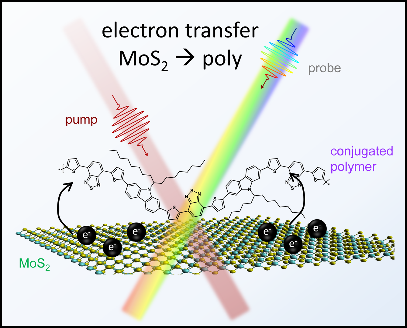
Femtosecond-transient absorption spectroscopy (fs-TAS) is an ultrafast technique that allows the investigation of excited state spectral dynamics [3]. Using fs-TAS, we studied the charge transfer dynamics of three organic/2D heterojunctions comprised of large-area monolayer MoS2 and solution-deposited semiconducting organic conjugated polymer thin-films (Figure 1). By changing the conjugated polymer from amorphous, disordered conjugated polymers, such as PCDTBT and PTB7, to more ordered, semi-crystalline conjugated polymers, such as P3HT, we showed that we can increase the lifetime of electrons transferred from 2D MoS2 by an order-of-magnitude. Due to their ease of fabrication and scalability, conjugated polymer/2D p-n heterojunctions can be considered good candidates for ultrathin electronic devices.
References Section 3.1 A:
[1] D. Jariwala, et al., Nature Mater. 16, 170 (2017).
[2] T. A. Shastry, et al., ACS Nano 10, 10573 (2016).
[3] J. Cabanillas-Gonzales, et al., Adv. Mater. 23, 5468 (2011).
B. Ultrafast control of the dimensionality of the exciton-exciton annihilation in atomically thin black phosphorus
[Physical Review Letters 124, 057403 (2020)]
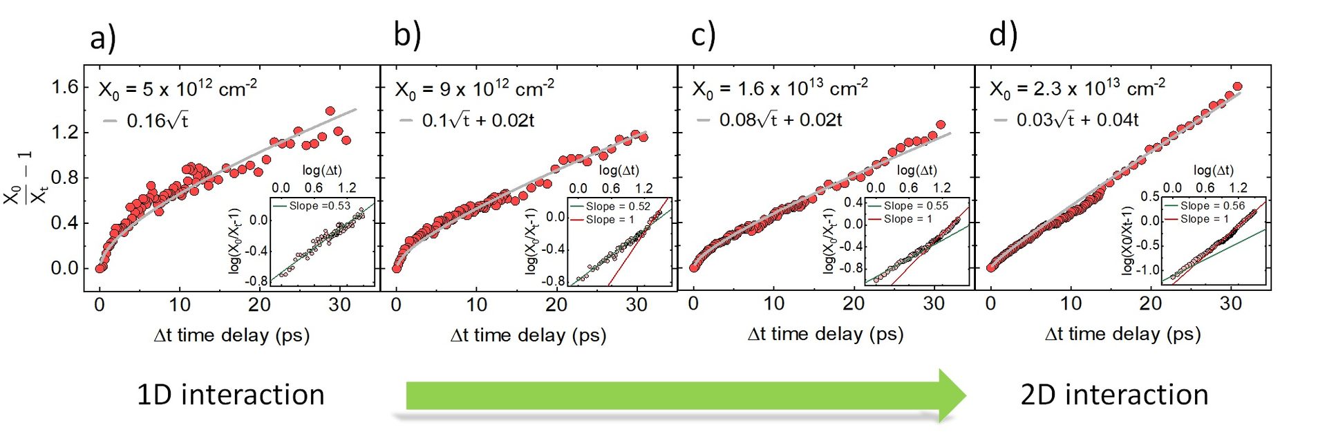
The dimensionality of a system dramatically influences its physical properties by altering the fundamental nature of electronic excitation and their interactions [1]. For example, in 1D systems (Carbon Nano Tubes – CNTs) the rate of exciton-exciton annihilation deviates from the mean filed approximation as the excitons are constrained by the physical dimensions to interact only with their nearest neighbors resulting in a time dependent rate [2]. However, in 2D (Transition metal dichalcogenides – TMDC) the dimensional restrictions are less stringent resulting in a time independent constant rate of annihilation consistent with the mean field approximation [3]. A particularly interesting case is presented by excitons in black phosphorus (BP), where quasi 1D excitons have been observed in atomically thin sheets of BP, resulting in a unique system of 1D excitons in a 2D plane. In this study, using micro- transient absorption spectroscopy(µ-TAS), we show that the interaction between excitons in atomically thin BP shows both 1D as well as 2D characteristics depending on the experimental parameters such as initial exciton density and temperature. We observe the classic 1D time dependent exciton-exciton annihilation dynamics at low exciton density (Figure 1a) and with increasing exciton density the data shows time independent rate, characteristic of a 2D (Figure 2d) exciton-exciton annihilation. We also observe more 1D characteristics at low temperature for all exciton densities. We attribute this effect to the anisotropic diffusion of excitons in atomically thin BP [4].
References Section 3.1 B:
[1] V. Gulbinas et al., J. Phys. Chem. 100(6), 2213 (1996).
[2] A. Srivastava et al., Phys. Rev. B 79, 205407 (2009).
[3] Y. Yu et al., Phys. Rev. B 93, 201111(R) (2016).
[4] V. Pareek et al., Phys. Rev. Lett. 124, 057403 (2020).
C. Investigation of nanoscale energy transport with time-resolved photoemission electron microscopy
[Chapter 10, Nanoscale Energy Transport : Emerging phenomena, methods and applications. Bristol, UK: Institute of Physics Publishing (2020)]
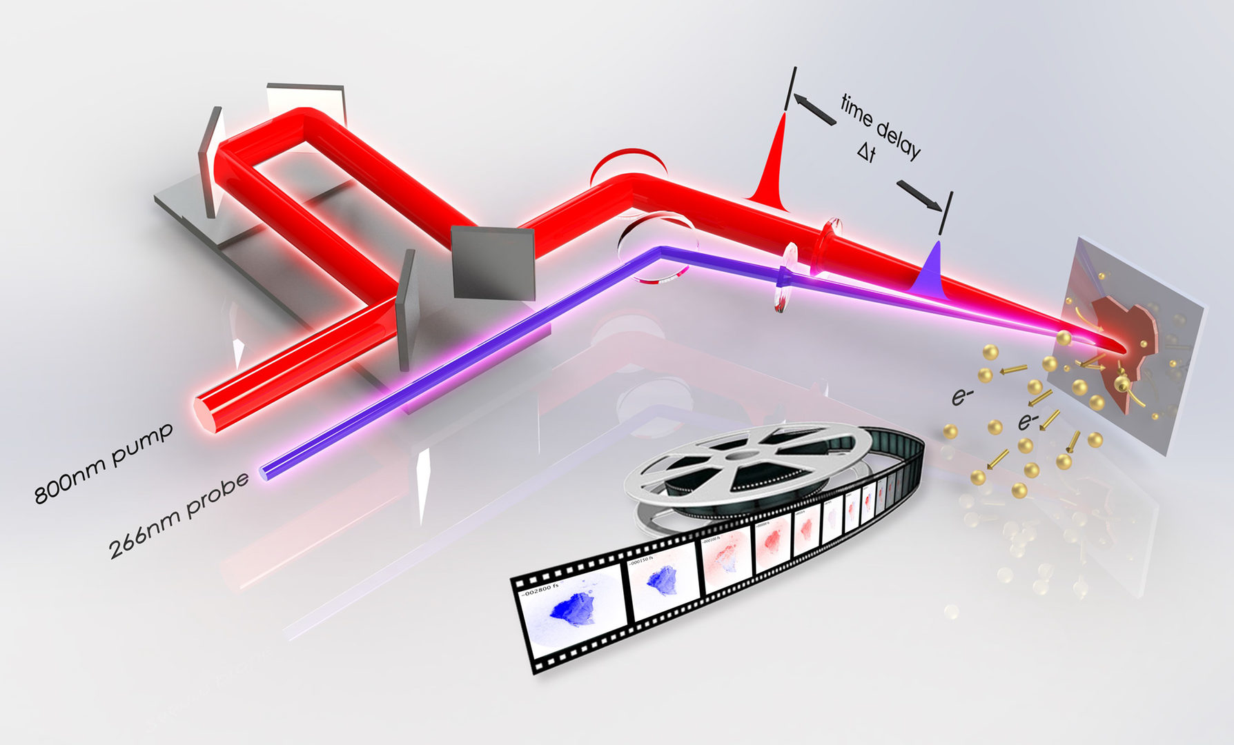
Electronics are becoming increasingly more compact and embedded with more functionality. Miniaturization of electronics leads to the emergence of new quantum phenomena, and the study of ultrafast electronic processes in these ultrasmall devices requires techniques that can simultaneously provide high spatial and temporal resolution. In response to this need, techniques such as ultrafast transient absorption microscopy, ultrafast electron microscopy, scanning ultrafast electron microscopy and time-resolved photoemission electron microscopy (TR-PEEM), have been developed.
The objective of this review is to elucidate electron microscopy techniques that have both high spatial and temporal resolution—with emphasis on the development and perspective of TR-PEEM techniques. In this review, we go through examples demonstrating the versatility of TR-PEEM in the study of carrier dynamics and charge transport in semiconductors [1-4], and we will look into the latest developments that promise to expand its capability, allowing TR-PEEM to explore electron dynamics and obtain detailed information, such as carrier scattering mechanisms and resolving the electronics spin texture.
References Section 3.1 C:
[1] M. K. L. Man et al., Nat. Nanotechnol. 12 36 (2017).
[2] E L. Wong, et al., Sci. Adv. 4 eaat9722 (2018).
[3] K. Fukumoto, et al., Appl. Phys. Lett. 104 053117 (2014).
[4] L. Wang, et al.. Nano Lett. 18 5172 (2018).
3.2 Femtosecond Techniques for Neurosciance Applications
A. Improving Signal and Photobleaching Characteristics of Temporal Focusing Microscopy with the Increase in Pulse Repetition Rate
[Methods and Protocols 2 (3), 65 (2019)]
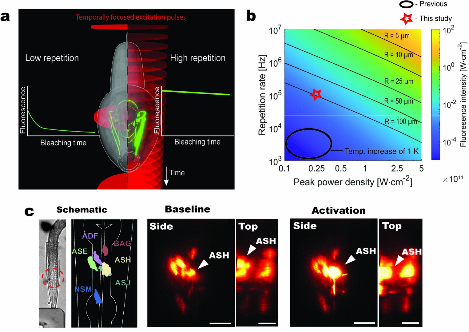
Wide-field temporal focused two-photon microscopy enables high-speed volumetric imaging via the simultaneous acquisition of a large sample area with high lateral and axial resolution [1,2]. Implementations to date have been held back due to the requirement for high fluence laser sources for excitation of a large area, which typically operated at a low repetition [3]. Achieving high signal intensity in this configuration necessitates increasing energy per pulse; however, this exacerbates the photobleaching in a power-law fashion. We demonstrate that the increase in the repetition rate of the laser system, within thermal constraints compatible with live imaging, strongly enhances fluorescence signal intensity without accelerating photobleaching (Fig. 4). We apply these findings to volumetric imaging of the nematode C. elegans sensory neurons. Recent advances in fiber lasers promise much more compact laser sources with full control of the repetition rate in a reliable and easy to operate package. Considering the advantages in speed offered by wide-field temporal focused two-photon microscopy, we envision that the new generation of fiber lasers will generate renewed and growing interest in temporal focusing technology.
References Section 3.2 A:
[1] D. Oron, et al., Opt. Express 13, 1468 (2005).
[2] G. Zhu, et al., Opt. Express 13, 2153 (2005).
[3] T. Schrödel, et al., Nat. Methods 10, 1013 (2013).
4. Publications
4.1 Journals
- C. E. Petoukhoff, S. Kosar, M. Goto, I. Bozkurt, M. Chhowalla, and K. M. Dani, Charge transfer dynamics in conjugated polymer/MoS2 organic/2D heterojunctions, Mol. Syst. Des. Eng. 4, 929 (2019).
- V. Lisicovas, B. M. K. Mariserla, C. Sahoo, R. T. Harding, M. K. L. Man, E. L. Wong, J. Madéo, and K. M. Dani, Improving signal and photobleaching characteristics of temporal focusing microscopy with the increase in pulse repetition rate, Methods Protoc. 2, 65 (2019).
- V. Pareek, J. Madéo, and K. M. Dani, Ultrafast control of the dimensionality of exciton-exciton annihilation in atomically thin black phosphorus, Phys. Rev. Lett. 124, 057403 (2020).
4.2 Books and Other One-Time Publications
- R. Wong, M. K. L. Man, and K. M. Dani, Investigation of nanoscale energy transport with time-resolved photoemission electron microscope., In B. Liao, Nanoscale Energy Transport Emerging phenomena, methods and application (pp.10-1 - 10-33). Bristol, UK: Institute of Physics Publishing (2020).
4.3 Oral and Poster Presentations
- Winchester, A. J., Macpherson, S., Pareek, V., Abdi-Jalebi, M., Andaji-Garmaroudi, Z., Petoukhoff, P., Wong, E., Madéo, J., Man, M. K. L., Stranks, S. D., Dani, K. M., Visualizing the Creation and Healing of Traps in Perovskite Photovoltaic Films by Light Soaking and Passivation Treatments, Oral Presentation, CLEO2019, San Jose, CA, USA May 05-10 (2019)
- Vivek, P., Madéo, J., Dani, K. M., 1D and 2D like exciton-exciton interaction in atomically thin black phosphorus, Oral Presentation, CLEO2019, San Jose, CA, USA May 05-10 (2019)
- Dani, K. M., Imaging the motion of charge with Time-Resolved Photoemission Electron Microscopy, Invited Talk, CLEO2019, San Jose, CA, USA May 05-10 (2019)
- Petoukhoff, C. E., Kosar, S., Bozkurt, I., Chhowalla, M., Dani, K. M., Charge carrier dynamics in conjugated polymer-MoS2 organic-2D heterojunctions, Poster Presentation, CLEO2019, San Jose, CA, USA May 05-10 (2019)
- Dani, K. M., Time resolved photoemission spectroscopies of semiconductor systems, Invited Talk, Fundamental Optical Processes in Semiconductors (FOPS) 2019, Banff, Canada, Aug 4-9 (2019)
- Urquizo, J. P., Madeo, J., Todorov, Y., Li, L., Davies, A. G., Linfield, E., Sirtori, C., Dani K. M., Patch Antenna Microcavities THz Quantum Cascade Lasers, Oral Presentation, IRMMW-THz, Paris, France, Sep 1-6 (2019)
- Urquizo, J. P., Madeo, J., Todorov, Y., Li, L., Davies, A. G., Linfield, E., Sirtori, C., Dani K. M., Patch Antenna Microcavities for THz Lasers, Oral Presentation, JSAP-OSA Joint Symposia- the 80th Autumm Meeting 2019, Sapporo, Japan Sep 18-21 (2019)
- Petoukhoff, C. E., Dani, K. M., O’Carroll, D. M., Ultrastrong Plasmon-Exciton Coupling between Ag Nanoparticles and Conjugated Polymers, Oral Presentation, JSAP-OSA Joint Symposia- the 80th Autumm Meeting 2019, Sapporo, Japan Sep 18-21 (2019)
- Urquizo, J. P., Madeo, J., Todorov, Y., Li, L., Davies, A. G., Linfield, E., Sirtori, C., Dani K. M., Hybrid patch-antenna microcavities for THz Quantum Cascade Lasers, Oral Presentation, Student Conference on Light OSA, Osaka, Japan, Nov (2019)
- Petoukhoff, C. E., Kosar, S., Bozkurt, I., Chhowalla, M., Dani, K. M., Spectroscopic Investigation of Electron Transfer from Monolayer MoS2 to Organic Conjugated Polymers, Oral Presentation, Materials research Society (MRS) Fall 2019, Boston, MA, USA Dec 1-6 (2019)
- Petoukhoff, C. E., Nonlinear and Ultrafast Optical Behavior of Mixed-Dimensional van der Waals Heterostructures, Invited Talk, Low Dimensional Materials for Optoelectronics (LDMO) 2019, Shenzhen, China, Dec 12-15 (2019)
- Petoukhoff, C. E., Kosar, S., Goto, M., Bozkurt, I., Chhowalla, M., Dani, K. M., Spectroscopic Investigation of Charge Transfer in Conjugated Polymer/MoS2 Organic/2D Heterojunctions, Oral Presentation, Progress in Electromagnetic Research (PIERS) 2019, Xiamen, China, Dec 17-20 (2019)
- Petoukhoff, C. E., Dani, K. M., O’Carroll, D. M., Ultrastrong Plasmon-Exciton Coupling between Ag Nanoparticles and Conjugated Polymers, Oral Presentation, Progress in Electromagnetic Research (PIERS) 2019, Xiamen, China, Dec 17-20 (2019)
5. Intellectual Property Rights and Other Specific Achievements
Nothing to report.
6. Meetings and Events
6.1 Seminar
- Date: April 22nd 2019
- Venue: C209 OIST Campus
- Speaker: Dr. Masahiko Tani
6.2 Workshop
Nothing to report.
7. Others
Nothing to report.