FY2018 Annual Report
Cellular and Molecular Synaptic Function Unit
Distinguished Professor (Fellow) Tomoyuki Takahashi

Abstract
In the presynaptic terminal, neurotransmitters are released from synaptic vesicles (SVs) via exocytosis. After exocytosis, SV membranes are retrieved by endocytosis and SVs are refilled with neurotransmitter to be reused for the next round of neurotransmission. This vesicular recycling/reuse mechanism is crucial for maintenance of synaptic transmission indispensable for brain functions. Although SV endocytic mechanisms are well studied recently, there is little study on the mechanism of vesicle transport after endocytosis. Using a giant synapse preparation in culture developped in our unit (Dimitrov et al, 2016 J Neurosci), we are real-time monitoring movements of SVs labeled with a fluorescent probe incorporated into them during endocytosis, analyzing trajectories and velocities of SV movements at resting terminals (Guillaud et al, 2017 eLife). We are now extending these techniques to monitor SV movements directed toward release sites in response to presynaptic fiber stimulation.
SV recycling is initiated by endocytosis. We found that the presynaptic protein α-synuclein, when over-loaded into presynaptic terminals from whole-cell pipettes, inhibits SV endocytosis (Eguchi et al, 2017 J Neurosci). This blocking effect could be rescued by co-loading microtubule (MT) depolymerizing drugs, suggesting that newly assembled MTs impair endocytosis. Endocytic inhibition by α-synuclein results in impairment of the fidelity of high-frequency neurotransmission that is required for a variety of brain functions. Since abnormal elevation of α-synuclein underlies Parkinson’s disease (PD), present results provide a synaptic model for PD. We extended these studies to tau protein toxicity underlying Alzheimer’s disease (AD). Loading tau into presynaptic terminals nearly abolished SV endocytosis, followed by a secondary impairment of exocytosis. Like α-synuclein, tau induced aberrant MT assembly, mimicked by taxol and its effect rescued by the MT-depolymerizing drug, nocodazole. Thus, α-synuclein and tau share a common mechanism of MT over-assembly, leading to impairments of precision and stability of fast neurotransmission.
Number of proteins reside on SVs and many more transiently interact with SVs. Our newly elaborated proteomics identified more than 1500 distinct SV-resident or interacting proteins. Using lenti-viral shRNA knockdown, we addressed functional roles of most abundant SV-interacting kinase, AaK1, and clarified that it plays significant roles in SV endocytosis, transport and fidelity of neurotransmission. We also discovered an entirely new SV-interacting protein, RDG1305455, having highly conserved primary structure within mammals. These proteome database will provide as a powerful tool for elucidating molecular synaptic mechanisms supporting brain functions.
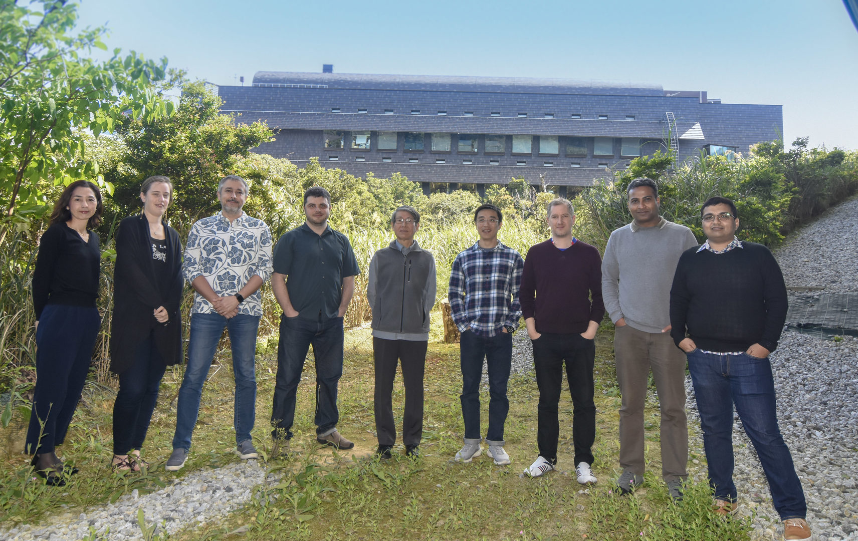
1. Staff
- Dr. Laurent Guillaud, Group Leader
- Dr. Zacharie Taoufiq, Staff Scientist
- Dr. Satyajit Mahapatra, Staff Scientist
- Dr. Han-Ying Wang, Postdoctoral Scholar
- Dr. Alemeh Zamani, Postdoctoral Scholar (until September 30, 2018)
- Dr. Soumyajit Dutta, Postdoctoral Scholar (since October 1, 2018)
- Dr. Dimitar Dimitrov, Specialist, Technical Staff
- Ms. Anna Garanzini, Technical Staff
- Mr. Francois Beauchain, Technical Staff
- Ms. Lashmi Piriya Ananda Babu, OIST Ph.D.Student (until May 31, 2018)
- Ms. Seiko Kawata, Research Intern (from August 1, 2018 to January 31, 2019)
- Ms. Sara Abdelaal, Lab Rotation Student (from September to December, 2018)
- Mr. Dvyne Nosaka, Lab Rotation Student (from Septembet to December, 2018)
- Ms. Sayori Gordon, Research Unit Administrator
2. Collaborations
2.1 Research theme: Regulatory mechanisms of transmitter release
- Name of partner organization: Doshisha University Faculty of Life and Medical Sciences
- Type of collaboration: Joint research
- Name of principal researcher: Dr. Tetsuya Hori, Doshisha University
- Name of researcher: Dr. Naoto Saitoh, Doshisha University
2.2 Research theme: Effect of photo-switchable microtubules disassembler PST on α-synclein-induced inhibition of vesicle endocytosis
- Name of partner organization: Department of Chemistry and Pharmacy and Centre for Integrated Protein Science, Ludwig-Maximillians-University
- Type of collaboration: Scientific Collaboration
- Name of principal researcher: Dr. Dirk Trauner
- Name of researcher: Dr. Oliver Thorn-Seshold
3. Activities and Findings
(1) Tau-induced tubulin assembly can impair synaptic transmission by blocking vesicle endocytosis. (Hori et al, manuscript in preparation)
Accumulation of soluble tau proteins causes neuronal dysfunctions leading to tauopathies, represented by Alzheimer’s disease (AD). When human recombinant tau was directly loaded into the calyx of Held presynaptic terminals in slice, EPSCs gradually declined in amplitude and reached ~50% in 30 min. This tau effect depended upon stimulation frequency, with stronger inhibition at higher frequencies. The microtubule (MT) assembler taxol mimicked these effects, whereas tau mutant lacking MT-binding site had no effect. Furthermore, the inhibitory effect of tau on EPSCs were fully rescued by co-loaded MT disassembler nocodazole. Finally, using immunofluorescence techniques with tubulin antibody, we confirmed that tau infused into presynaptic terminals significantly increased MT assembly. In membrane capacitance recording from calyceal presynaptic terminals, tau loading inhibited SV endocytosis, followed by an inhibition of SV exocytosis, with a delay of ~10 min. The inhibitory effect of tau on SV endocytosis was also rescued by nocodazole. We conclude that accumulation of soluble tau leads to gain of toxic function via de novo MT assembly, inhibiting SV endocytosis and recycling, thereby impairing high frequency neurotransmission. These mechanisms of tau toxicity are like those of α-synuclein (Eguchi et al, 2017, J Neurosci) suggesting that a common therapeutic tool might potentially be elaborated for both PD and AD.
(2) Volatile anesthetic isoflurane inhibits high-frequency excitatory neurotransmission via direct inhibition of vesicle fusion (Wang et al, manuscript in preparation)
Surprisingly little is known for the mechanism of volatile anesthesia. Bath-application of the anesthetic isoflurane, at clinical doses, attenuated neurotransmission at the calyx of Held in slices, by reducing release probability and number of functional release sites. In presynaptic whole-cell recordings, we clarified that isoflurane inhibits voltage-gated Ca2+ channels that play a triggering role in transmitter release. In presynaptic capacitance measurements, we found that isoflurane directly inhibits SV exocytosis in response to a sustained Ca2+ influx. In simultaneous pre- and postsynaptic AP recordings, isoflurane impaired synaptic fidelity, preferentially during high-frequency transmission. Because high-frequency transmission is associated with sustained Ca2+ influx and plays pivotal roles in various brain functions, we conclude that inhibition of exocytic fusion machinery by isoflurane contributes most importantly to its anesthetic actions.
(3) Astrocytic NO drives presynaptic cascade for inducing ischemic long-term potentiation at hippocampal CA1 synapses (Takagi et al, unpublished)
At hippocampal CA1 region in slice, oxygen/glucose deprivation (OGD) induces ischemic long-term potentiation (iLTP), associated with a decrease in paired-pulse ratio, a conventional index suggesting an increase in transmitter release. Pharmacological tests confirmed involvement of NMDA receptors and NO cascade in iLTP. Further pharmacological dissections revealed that the downstream of NO involves PKG, Rho, Rho kinase and PIP2. Thus, amazingly, the mechanisms likely overlap with those underlying exo-endocytic SV balancing at the calyx of Held (Eguchi et al, 2012, Neuron). Distinctly, however, a CamKII inhibitor attenuated iLTP. Since CaMKII inhibits neuronal NO synthase, but activates astrocytic epithelial NOS, it is speculated that OGD releases NO from astrocytes, thereby activating the presynaptic NO cascade for SV recycling enhancement. We are to test this hypothesis by destroying astrocytes using fluoroacetate.
(4) Hidden proteome of synaptic vesicles (Taoufiq et al, manuscript in preparation)
SV fraction was purified from adult rat brain, and multi-step enzymatic digestions followed by multi-dimensional peptide separation were performed. These enabled comprehensive proteomics (UD proteomics) and revealed ~1500 proteins in SV fraction. These SV proteins were quantified for their abundance, copy number per SV, residing ratio at SV relative to other presynaptic compartments. This SV proteome also revealed whole SV protein subfamilies and subunits, many of which remained undetected in previous SV proteomics. Among SV-interacting kinases, AaK1was found to be most abundant. Functional analyses, using lentiviral shRNA knockdown, in combination with pHluorin imaging, and patch-clamp recording, revealed that AaK1 accelerates SV endocytosis and transport, thereby supporting fidelity of high-frequency neurotransmission.
(5) Functional roles of cytoskeletal elements in presynaptic terminals (Ananda Babu et al, manuscript in preparation)
Presence of MT in nerve terminals is controversial and its functional role is totally unknown. At the calyx of Held, STED microscopy revealed that MTs extend from axon into the depth of presynaptic terminals where SVs are present. After live imaging of MTs using SiR-tubulin loaded into the calyceal terminals by incubation, the MT disassembler vinblastine, incubated with slices, gradually depolymerized MTs thereby reducing SiR-tubulin fluorescence intensity. After 20 min incubation with vinblastine, which depolymerized MTs by ~30 %, EPSCs evoked by a train of stimuli (100 Hz x30) at the calyx of Held underwent a short-term depression (STD) followed by bi-exponential recovery, with a sub-second fast time constant(tf) followed by a second-order slow time constant (ts). The 20-min vinblastine treatment had no effect on the tf but markedly prolonged the ts. In marked contrast, the F-actin disassembler, latrunculin A, specifically inhibited the tf with no effect on the tf Co-treatment of vinblastine and latrunculin A prolonged both tf and tf, with no sign of mutual interference. Both vinblastine and latrunculin impaired the fidelity of high-frequency neurotransmission, with the effect of former stronger than the latter. We conclude that MTs and F-actin in presynaptic terminals play essential and distinct roles in SV transport, thereby contributing to the maintenance of high-frequency neurotransmission.
(6) Regulatory movements of synaptic vesicles in calyx-type synapses (Ohmachi et al, manuscript in preparation)
During neurotransmission, SVs are transported to release sites in nerve terminals, but such SV movements are not sufficiently characterized. Quantum (Q) dot, unconjugated with SV protein, were scarcely loaded into cultured calyceal presynaptic terminals via SV endocytosis to follow single SV movements in calyceal terminals. SV mobility at resting terminals was low and random, but during repetitive stimulation, it became faster and directional. After a sudden increase in mobility, SV fluorescence signal sometimes vanishes. Since Q-dot fluorescence does not bleach during recording and average SV displacement during 1-s sampling time is much smaller than the spatial resolution of confocal optics, the fluorescence vanishment is most likely caused by exocytic release of Q dot. In 9 such examples, there was a positive correlation between the travel distance and time to vanish, indicating nearly constant SV movements at 50 nm/s. Furthermore, the travel distance was proportional to the latency of uni-directional SV movements after stimulus onset. These results suggest that the SV transport mechanism directed to release sites is unique. They also suggest that a putative triggering factor, which initiates directional movements of SVs may derive from a location not far from release sites.
4. Publications
4.1 Journals
Yamashita M, Kawaguchi S, Hori T, Takahashi T (2018). "Vesicular GABA uptake can be rate limiting for recovery of IPSCs from synaptic depression". Cell Reports 22, 3134-3141.
4.2 Books and other one-time publications
Nothing to Report.
4.3 Oral and Poster Presentations
Oral Presentations
Takahashi, T. “Excitatory and inhibitory neurotransmitter refilling into synaptic vesicles”. NIPS Research Meeting: New developments in multi-layer functional analysis based on visualization and manipulation of signaling dynamics. National Institute for Physiological Sciences, Aichi. September 27- 28, 2018.
Taoufiq, Z. “Exploring the Hidden Proteome of Synaptic Vesicles”. JSPS Core-to-Core program “Nanobiology of neural plasticity based on optical nanoscopy” Symposium 2018. Doshisha University, Kyoto. December 1-3, 2018.
Guillaud, L. “ATP-dependent phase transition of peri-active zone and active zone bio-condensates regulate synapse organization and function”. ASCB/EMBO 2018 meeting. San Diego Convention Center, CA, U.S.A., December 8 - 12, 2018
Poster Presentations
Guillaud, L. and Takahashi, T. “Mitochondrial activity-dependent phase transitions of pre-synaptic bio-condensates regulate synchronous trafficking of synaptic vesicles and AMPA receptors in giant mammalian synapses”. Gordon Research Conference: Cell Biology of the Neuron. Waterville Valley, U.S.A. June 24 - 29, 2018.
Taoufiq, Z. “The Hidden Proteome of Synaptic Vesicles underlies Brain Disease Phenocopies”. Gordon Research Seminar: Molecular and Cellular Neurobiology "Molecular and Cellular Dynamics of Neural Function".
Two types of work: Discussion Leader in the 'Neural Cell Connectivity and Disease' session and Poster presentation. The Hong Kong University of Science and Technology, Hong Kong, China. June 30 - July 1, 2018.
Taoufiq, Z., Ninov, M., Jahn, R., Takahashi, T. “The Hidden Proteome of Synaptic Vesicles underlies Brain Disease Phenocopies”. Gordon Research Conference: Molecular and Cellular Neurobiology "Recent Discoveries and Technological Advances in the Study of the Nervous System during Development, Health and Disease". The Hong Kong University of Science and Technology, Hong Kong, China. July 1- 6, 2018.
Wang, H-Y., Eguchi, K., Takahashi, T. “Isoflurane preferentially inhibits high-frequency excitatory neurotransmission via directly inhibiting vesicle fusion mechanism”. Neuroscience 2018, San Diego Convention Center, CA, U.S.A. November 3- 7, 2018
Guillaud, L. and Takahashi, T. “ATP-dependent liquid phase transitions of presynaptic bio-condensates regulate synapse organization and function”. The OIST mini symposium - The 16th international membrane research forum. OIST Campus, Lab 3, March 18, 2019
Taoufiq, Z., Wang, H-Y. Villar-Briones, A. Beauchain, F. Sasaki, T. Takahashi, T. "Anchored Protein Complexes at Synaptic Vesicular Membrane and Neurotransmission Computation." The OIST mini symposium-The 16th international membrane research forum. OIST Campus, Lab3, March 18, 2019
5. Intellectual Property Rights and Other Specific Achievements
Nothing to report
6. Meetings and Events
6.1 Invited Seminar
- Name of the Workshop: Developmental Neurobiology Course 2018
- Date: August 6, 2018
- Venue: OIST Center bldgs.
- Speaker: Prof. Tomoyuki Takahashi
- Lecture title: Recycling reuse of synaptic vesicles.
~ Panel discussion: Dr. Erwin Neher and Prof. Takahashi
- Name of the Conference: The OIST mini symposium - The 16th international membrane research forum
- Date: March 20, 2019
- Venue: OIST Center bldgs.
- Speaker: Prof. Tomoyuki Takahashi
- Lecture title: Transport of synaptic vesicles in mammalian nerve terminals.
- Name of the Conference: Neuroscience conference for Parkinson Disease - Second series
- Date: March 22, 2019
- Venue:Okinawa Medical Association
- Speaker: Prof. Tomoyuki Takahashi
- Lecture title: Recycling reuse of synaptic vesicles and neuronal diseases.
6.2 OIST Seminar, host by Prof. Takahashi
- Date: April 13, 2018
- Venue: OIST Campus, Lab1
- Speaker: Dr. Kazuhiko Yamaguchi, RIKEN Brain Science Institute
- Talk title: Reassessment of mechanism underlying cerebellar learning.
7. Other
7.1 Teaching class ‘Introduction to super resolution microscopy (nanoscopy)
- Imaging beyond the diffraction limit’, part of the 'Emerging technologies in life
sciences' course. For the first year PhD students organized by Pr. Ichiro Maruyama, OIST - Date: July 9, 2018
- Lecturer: Laurent Guillaud


