FY2022 Annual Report
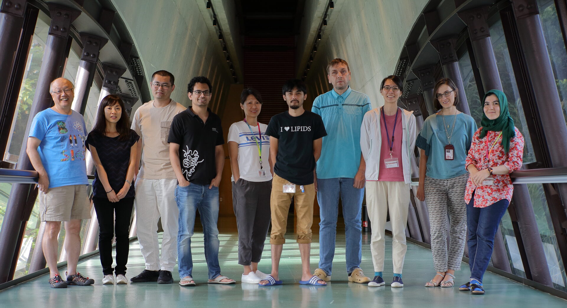
Membrane Cooperativity Unit
Professor Akihiro Kusumi

Abstract
We at the Membrane Cooperativity Unit are working hard to reveal how the dynamic platforms for signal transduction and the synapses for the neuronal transmission form and function in the plasma membrane. For this purpose, we take a unique approach (in addition to other more conventional approaches). Namely, we develop new and unique methods for single-molecule imaging and manipulation at nanometer precisions in living cells, with a special attention paid to high time resolutions (world’s fastest single fluorescent-molecule imaging). The smooth liaison between physics/engineering and biomedicine is a key for our research.
The plasma membrane is the outermost membrane of the cell, and thus it encloses the entire cell. It is critically important for the cell - the fundamental unit of life - because it defines the space for it. The plasma membrane exchanges information, energy, and substances with the outside world, and we pay special attention to the mechanism for signal transfer from outside to inside the cell, a function generally called “signal transduction”. In the signal transduction process, the plasma membrane works like a sensor + computer + effector.
The Membrane Cooperativity Unit strives to understand how the plasma membrane works at very fundamental levels, based on unique insights we obtain by applying single-molecule imaging-tracking methods. More specifically, we are now revealing the mechanisms by which the metastable molecular complexes and meso-scale membrane domains, including membrane compartments, raft domains, and protein oligomers, form and work in concert to enable signal transduction and synapse formation/modulation in/on the plasma membrane.
1. Staff
- Dr. Amine Betul Nuriseria Aladag, Post Doctoral Scholar
- Dr. HooiCheng Lim, Post Doctoral Scholar
- Dr. Bo Tang, Post Doctoral Scholar
- Dr. Taka-Aki Tsunoyama, Post Doctoral Scholar
- Dr. Maoji Wang, Post Doctoral Scholar
- Dr. Peng Zhou, Post Doctoral Scholar
- Dr. Jun-Seok Lee, Technician
- Dr. Irina Meshcheryakova, Technician
- Ms. Yuri Nemoto, Technician
- Mr. Ryuto Shinoaki, Technician
- Mr. Saahil Acharya, PhD Student
- Mr. Jerome Theodore Tinker, Rotation Student
- Ms. Sharon Babar, Research Intern
- Ms. Takako Dakeshita, Research Intern
- Mr. Fuyuki Higa, Research Assistant (Part-time)
- Mr. Yusuke Higa, Research Assistant (Part-time)
- Mr. Yuta Kogi, Research Assistant (Part-time)
- Mr. Rin Nakama, Research Assistant (Part-time)
- Mr. Yoshito Takaesu, Research Assistant (Part-time)
- Mr. Ryu Tsuha, Research Assistant (Part-time)
- Ms. Sachie Matsuoka, Research Unit Administrator
- Dr. Akihiro Kusumi, Professor
2. Collaborations
2.1 Revealing the dynamics, structure, and function of metastable signaling molecular complexes by single-molecule imaging
- Description: Developing ultrafast 3D single-molecule imaging, and applying it to revealing the dynamics and formation mechanism of the signaling complex in synaptic signaling, Fcepsilon signaling, focal adhesion architecture and signaling, and GPI-anchored proteins’ raft-based signaling
- Type of collaboration: Joint research
- Researchers:
- Dr. Takahiro Fujiwara, Associate Professor, Institute for Integrated Cell-Material Sciences (iCeMS), Institute of Advanced Studies, Kyoto University
- Dr. Kenichi Suzuki, Professor, G-CHAIN, Gifu University
2.2 Unraveling the large-scale molecular-species selective diffusion barriers in the axonal initial segment in the neuron using ultrafast single-molecule imaging
- Description: By applying ultrafast single-molecule imaging and ultrafast single-molecule localization microscopy developed by us, we try to unravel the large-scale molecular-species selective diffusion barriers in the axonal initial segment in the neuron
- Type of collaboration: Joint research
- Researchers:
- Dr. Takahiro Fujiwara, Associate Professor, Institute for Integrated Cell-Material Sciences (iCeMS), Institute of Advanced Studies, Kyoto University
2.3 Revealing super-transient signaling molecular complexes by single-molecule imaging and tracking
- Description: To reveal the mechanisms for the formation and function of super-transient signaling molecular complexes using single-molecular imaging and tracking
- Type of collaboration: Joint research
- Researchers:
- Dr. Takahiro Fujiwara, Associate Professor
- Dr. Hisae Tsuboi, Resercher
Institute for Integrated Cell-Material Sciences (iCeMS), Institute of Advanced Studies, Kyoto University
2.4 Elucidation of dynamics and formation mechanisms of cellular signaling complexes by developing new single particle tracking methods
- Description: Developing fluorescent probes for their applications to single-molecule imaging in living cells, and by using the developed probes, elucidating dynamics and formation mechanisms of cellular signaling complexes induced by various intercellular signaling molecules and alien antigens, including (non-pathogenic) viruses
- Type of collaboration: Joint research
- Researchers:
- Dr. Dai-Wen Pang, Professor
- Dr. An-An Liu, Lecturer
- Ms. Dan-dan Fu, PhD candidate
Department of Chemistry, College of Chemistry, Nankai University,P. R. China
2.5 Elucidating the functions of plasma membrane compartmentalization
- Description: Elucidating how the signal transduction functions of the plasma membrane is regulated using the actin-based compartmentalization of the plasma membrane, using ultrafast single-molecule imaging-tracking and super-resolution microscopy
- Type of collaboration: Joint research
- Researchers:
- Dr. Pakorn Tony Kanchanawong, Professor
Mechanobiology Institute, The National University of Singapore
- Dr. Pakorn Tony Kanchanawong, Professor
2.6 Development of deep-learning methods for single-molecule imaging experiments and analysis
- Description: Developing AI-based methods for performing single-molecule imaging and for analyzing single-molecule imaging data
- Type of collaboration: Joint research
- Researchers:
- Dr. Kazuhiro Hotta, Professor
Mechanobiology Institute, Department of Electrical and Electronic Engineering, Faculty of Engineering, Meijo University
- Dr. Kazuhiro Hotta, Professor
2.7 Developing single-molecule imagining hardware to be used for AI-based single-molecule imaging and analysis software
- Description: Developing software and hardware for automating single fluorescent-molecule observations in living cells and data analysis
- Type of collaboration: Joint research
- Researchers:
- Dr. Kazuhiro Hotta, Professor
Mechanobiology Institute, Department of Electrical and Electronic Engineering, Faculty of Engineering, Meijo University
- Dr. Kazuhiro Hotta, Professor
2.8 Revealing the mechanisms for the synapse formation and long-term potentiation by combining super-resolution microscopy and single-molecule imaging
- Description: To discover the mechanisms for functional and structural synaptic plasticity underlying learning and memory, by the combined use of super-resolution microscopy and single-molecule imaging
- Type of collaboration: Joint research
- Researchers:
- Dr. Michisuke Yuzaki, Professor
Graduate School of Medicine, Keio University
- Dr. Michisuke Yuzaki, Professor
2.9 Elucidation of nano-scale localization of synaptic molecules in living neurons of mice
- Description: To develop a platform for correlative live single-molecular imaging and super-resolution microscopy and optimize it, based on the feedback from Keio
- Type of collaboration: Joint research
- Researchers:
- Dr. Michisuke Yuzaki, Professor
- Dr. Kazuya Nozawa, Assistant Professor
- Dr. Tetsuko Fukuda, Researcher
Graduate School of Medicine, Keio University
2.10 Revealing the cellular signaling platforms formed by regulated liquid-liquid phase separation(LLPS)
- Description: To reveal the mechanisms by which nano-micron-sized liquid-like signaling platforms form and facilitate signaling in the cell and between the cells
- Type of collaboration: Joint research
- Researchers:
- Dr. Dragomir Milovanovic, Lab Head
- Dr. Christian Hoffmann, Postdoctoral Fellow
- Mr. Gerard Aguilar Perez, PhD Student
DZNE; German Center for Neurodegenerative Diseases
3. Activities and Findings
3.1 Discovering the critical roles for EGFR and EGFR-HER2 clusters in EGF binding
Epidermal growth factor (EGF) signaling regulates normal epithelial and other cell growth, with EGF receptor (EGFR) overexpression reported in many cancers. However, the role of EGFR clusters in cancer and their dependence on EGF binding is unclear. We, in collaboration with Prof. M. C. Leake and his colleagues at the University of York, performed novel single-molecule total internal reflection fluorescence microscopy of (i) EGF and EGFR in living cancer cells, (ii) the action of anti-cancer drugs that separately target EGFR and human EGFR2 (HER2) on these cells and (iii) EGFR-HER2 interactions. We selected human epithelial SW620 carcinoma cells for their low level of native EGFR expression, for stable transfection with fluorescent protein labelled EGFR, and imaged these using single-molecule localization microscopy to quantify receptor architectures and dynamics upon EGF binding. Prior to EGF binding, we observe pre-formed EGFR clusters. Unexpectedly, clusters likely contain both EGFR and HER2, consistent with co-diffusion of EGFR and HER2 observed in a different model CHO-K1 cell line, whose stoichiometry increases following EGF binding. We observe a mean EGFR : EGF stoichiometry of approximately 4 : 1 for plasma membrane-colocalized EGFR-EGF that we can explain using novel time-dependent kinetics modelling, indicating preferential ligand binding to monomers. Our results may inform future cancer drug developments.
3.2 Revealing an important function of sphingomyelin-sequestered cholesterol domain in the plasma membrane: Sphingomyelin-sequestered cholesterol domain recruited/produced at clathrin-coated pits recruits formin-binding protein 17 for constricting clathrin-coated pits in influenza virus entry
Influenza A virus (IAV) is a global health threat. The cellular endocytic machineries harnessed by IAV remain elusive. By tracking single IAV particles and quantifying the internalized IAV, we, in collaboration with Prof. Dai-Wen Pang and his colleagues at Nankai University in China, found that sphingomyelin (SM)-sequestered cholesterol, but not accessible cholesterol, is essential for the clathrin-mediated endocytosis (CME) of IAV. The clathrin-independent endocytosis of IAV is cholesterol independent, whereas the CME of transferrin depends on SM-sequestered cholesterol and accessible cholesterol. Furthermore, three-color single-virus tracking and electron microscopy showed that the SM-cholesterol complex nanodomain is recruited to the IAV-containing clathrin-coated structure (CCS) and facilitates neck constriction of the IAV-containing CCS. Meanwhile, formin-binding protein 17 (FBP17), a membrane-bending protein that activates actin nucleation, is recruited to the IAV-CCS complex in a manner dependent on the SM-cholesterol complex. We propose that the SM-cholesterol nanodomain at the neck of the CCS recruits FBP17 to induce neck constriction by activating actin assembly. These results unequivocally show the physiological importance of the SM-cholesterol complex in IAV entry.
3.3 Live-cell imaging of single neurotrophin receptor molecules on human neurons in Alzheimer’s disease
Neurotrophin receptors such as the tropomyosin receptor kinase A receptor (TrkA) and the low-affinity binding p75 neurotrophin receptor p75NTR play a critical role in neuronal survival and their functions are altered in Alzheimer's disease (AD). Changes in the dynamics of receptors on the plasma membrane are essential to receptor function. However, whether receptor dynamics are affected in different pathophysiological conditions is unexplored. Using live-cell single-molecule imaging, we, in collaboration with the late Prof. Istvan Abraham and his team members at the University of Pécs Medical School, examined the surface trafficking of TrkA and p75NTR molecules on live neurons that were derived from human-induced pluripotent stem cells (hiPSCs) of presenilin 1 (PSEN1) mutant familial AD (fAD) patients and non-demented control subjects. Our results show that the surface movement of TrkA and p75NTR and the activation of TrkA- and p75NTR-related phosphoinositide-3-kinase (PI3K)/serine/threonine-protein kinase (AKT) signaling pathways are altered in neurons that are derived from patients suffering from fAD compared to controls. These results provide evidence for altered surface movement of receptors in AD and highlight the importance of investigating receptor dynamics in disease conditions. Uncovering these mechanisms might enable novel therapies for AD.
4. Publications
4.1 Journals
Original Articles
- A. J. M. Wollman, C. Fournier, I. Llorente-Garcia, O. Harriman, A. L. Payne-Dwyer, S. Shashkova, P. Zhou, T.-C. Liu, D. Ouaret, J. Wilding, A. Kusumi, W. Bodmer, and M. C. Leake. Critical roles for EGFR and EGFR-HER2 clusters in EGF binding of SW620 human carcinoma cells. J. Royal Society Interface 20220088 (2022) doi: 10.1098/rsif.2022.0088
- B. Tang, E.-Z. Sun, Z.-L. Zhang, S.-L. Liu, J. Liu, A. Kusumi, Z. Hu, T. Zeng, Y.-F. Kang, H.-W. Tang, and D.-W. Pang. Sphingomyelin-sequestered cholesterol domain recruits formin-binding protein 17 for constricting clathrin-coated pits in influenza virus entry. J. Virol. 96: e01813-21 (2022). doi: 10.1128/jvi.01813-21
- K. Barabás, J. Kobolák, S. Godó, T. Kovács, D. Ernszt, M. Kecskés, C. Varga, T. Z. Jánosi, T. Fujiwara, A. Kusumi, A. Téglási, A. Dinnyés, and I. M. Abraham. Live-cell imaging of single neurotrophin receptor molecules on human neurons in Alzheimer’s disease. Int. J. Mol. Sci. 22: 13260 (2022). doi: 10.3390/ijms.222413260
Review Articles
- K. G. N. Suzuki and A. Kusumi. Refinement of Singer-Nicolson fluid-mosaic model by microscopy imaging: Lipid rafts and actin-induced membrane compartmentalization. Biochim. Biophys. Acta - Biomembranes 1865, 184093 (2023). doi:10.1016/j.bbamem.2022.184093
4.2 Books and other one-time publications
Nothing to report
4.3 Oral and Poster Presentations
Oral Presentations
-
A. Kusumi. Metastable signaling platforms as revealed by single-molecule imaging:
mutual interest between István and me. The Second symposium on super-resolution and advanced fluorescence microscopy and István Ábrahám memorial workshop. Pécs, Hungary. April 1, 2022 (Remote Talk). -
A. Kusumi. Metastable nano-liquid signaling platforms on the cell membrane as revealed by single-molecule imaging. Opening Plenary Lecture. The 17th IEEE International Conference on Nano/Micro Engineered and Molecular Systems (IEEE MEMS 2022) Virtual Meeting in Taipei (Taiwan) and Los Angeles (U.S.A.). April 14, 2022.
-
A. Kusumi. Development of the ultrafast camera for single-molecule imaging and detection of the plasma membrane compartmentalization. OIST Symposium: Cells, Energetics, and Information: New Perspectives on Nonequilibrium Systems. Okinawa. June 9, 2022
-
A. Kusumi. Development of the ultrafast camera system for single-molecule imaging and metastable nano-liquid signaling platforms on the cell membrane. OIST-Kyoto University Joint Workshop: Challenge in Biomedical Complexity. Okinawa. November 3, 2022.
-
A. Kusumi. Development of the ultrafast camera system for single-molecule imaging and discovery of metastable nano-liquid signaling platforms on the cell membrane. OIST Workshop: Recent Trends in Microrheology and Microfluidics. Okinawa. January 12, 2023.
-
A. Kusumi. Single-molecule imaging studies of postsynaptic receptor turnover on the PSD protein condensates. 15th NeuroWissenschaftliche Gesellshaft (German Neuroscience Society) Meeting. Symposium 23: Phase separation in neuronal (patho)physiology, Göttingen, Germany. March 24, 2023.
-
T. Tsunoyama. Nanoscale condensed liquid platform on the plasma membrane for signal integration. OIST-Kyoto University Joint Workshop: Challenge in Biomedical Complexity. Okinawa. November 3, 2022.
-
T. Tsunoyama. Nanoscale LLPS-based liquid-like signaling platform that cooperatively integrates RTK, GPCR, and GPI-anchored receptor signals. The 45th Annual Meeting of the Molecular Biology Society of Japan (MBSJ). Makuhari, Chiba. December 1, 2022.
Poster Presentations
-
S. Acharya. Syngap LLPS condensates as the basic platform for recruiting PSD95 and receptor oligomers for generating neuronal excitatory synapses. Biophysical Society 67th Annual Meeting. San Diego, U.S.A. February 21, 2023.
5. Intellectual Property Rights and Other Specific Achievements
Japanese Patent Application
Application Number: 2022-096238
Application Date: 15 June 2022
Title of Application:
Peptide drugs for suppressing tolerance development by blocking homo- and
hetero-dimerization of opioid receptors
6. Meetings and Events
Nothing to report
7. Other
Nothing to report


