

Mustafa Sami, Ph.D.
Email: m.sami(at)oist.jp
Professional Experience
- OIST Graduate University (Technician)
- RIKEN Center for Biosystems Dynamics Research, BDR (Research Scientist)
- RIKEN Center for Developmental Biology, CDB (Technician)
- Kobe Institute of Computing, KIC, (Instructor)
- Nippon Boehringer Ingelheim Co., LTD. (Research Fellowship)
- Tampere University of Technology, TUT, (Exchange Researcher)
- Niigata University (Research Fellowship)
Research Interest
Signal Processing, Digital Image Processing, Complex Network Analysis, Deep Learning, Machine Learning, Pattern Recognition, Quantitative Biology, Computer-Aided Diagnosis, Image Segmentation, Feature Selection and Extraction, Color Processing, GUI Development, Video and Text Processing and Retrieval.
Teaching Experience
- Digital Image Processing for Quantitative Biology, RIKEN Kobe
- Image Processing Using MATLAB, RIKEN Kobe
- Using AI for Clinical Applications, Kobe Institute of Computing (KIC)
Research Activities
Cell and Developmental Biology
- Leading the development of image-based software systems and GUI applications to process and analyze confocal microscopic images. Our goal was to understand early cell shape changes during organ growth and development in Drosophila. These images are often noisy and challenging to extract important features from. To address this, we designed specific intensity and morphological filters to help biologists observe and track cells automatically.
- Mustafa M. Sami, Yosuke Ogura, Yu-Chiun Wang, and Shigeo Hayashi, “Automated image processing and analysis software for epithelial cells quantification” 17th International European Light Microscopy Initiative Meeting (ELMI 2017), Dubrovnik, Croatia, May 2017.
- Michiko Takeda, Mustafa M. Sami, Yu-Chiun Wang, A homeostatic apical microtubule network shortens cells for epithelial folding via a basal polarity shift, Nature Cell Biology, Vol.20, pp. 36-45, Dec.2017.
- Yosuke Ogura, Fu-Lai Wen, Mustafa M. Sami, Tatsuo Shibata, Shigeo Hayashi, A Switch-like Activation Relay of EGFR-ERK Signaling Regulates a Wave of Cellular Contractility for Epithelial Invagination. Developmental Cell, Vol.46, pp. 162-172, July 2018.
- Anthony S Eritano, Claire L Bromley, Antonio Bolea Albero, Lucas Schütz, Fu-Lai Wen, Michiko Takeda, Takashi Fukaya, Mustafa M. Sami, Tatsuo Shibata, Steffen Lemke, and Yu-Chiun Wang. Tissue-scale mechanical coupling reduces morphogenetic noise to ensure precision during epithelial folding. Developmental Cell, Vol.53, pp. 212–228, April 2020.
- Leading the development of an automated software system to quantify fluorescence energy transfer (FRET) signals for activated extracellular signal-regulated kinase (ERK). This system was used to monitor the spatio-temporal dynamics of ERK during neuroectoderm patterning in Drosophila embryos.
- Yosuke Ogura#, Mustafa M. Sami#, Housei Wada, Shigeo Hayashi, Automated FRET quantification reveals distinct subcellular ERK activation kinetics in response to graded EGFR signaling in Drosophila. Genes to Cells, Vol.24, pp. 297–306, Feb. 2019. (# contributed equally)
- Leading the image processing and analysis of Electron Microscopy (EM) images for the nanopore formation project, focusing on the cuticle of the insect olfactory sensillum system.
- Toshiya Ando, Sayaka Sekine, Sachi Inagaki, Kazuyo Misaki, Laurent Badel, Hiroyuki Moriya, Mustafa M. Sami, Yuki Itakura, Takahiro Chihara, Hokto Kazama, Shigenobu Yonemura, Shigeo Hayashi. Nanopore Formation in the cuticle of an insect olfactory sensillum. Current Biology, Vol.29, pp. 1–9, May 2019.
- Leading the development of a quantification method for super-resolution live imaging of super-cellular circumferential actin cable formation during tracheal tubulogenesis.
- Sayaka Sekine, Mustafa M. Sami, Housei Wada, Shigeo Hayashi, “Super-resolution live imaging of supercellular circumferential actin cable formation during tracheal tubulogenesis”, Joint Annual Meeting of JSDB and JSCB, Tokyo, Jun. 2018.
- Sayaka Sekine, Mitsusuke Tarama, Housei Wada, Mustafa M. Sami, Tatsuo Shibata & Shigeo Hayashi, "Emergence of periodic circumferential actin cables from the anisotropic fusion of actin nanoclusters during tubulogenesis", Nature Communications, Jan. 2024.
Heart-on-a-chip Micro Device
- Leading the development of a software tool to track nano-bead particles for a new device that generates heart tissues from human iPS cells. This tool can be used for drug discovery for heart diseases and cardiac toxicity tests. Tracking these nano-beads is challenging due to their small size and irregular speed movements in complex tissue environments.
- Mosha Abulaiti, Yaxiaer Yalikun, Kozue Murata, Asako Sato, Mustafa M. Sami, Yuko Sasaki, Yasue Fujiwara, Kenji Minatoya, Yuji Shiba, Yo Tanaka & Hidetoshi Masumoto. Establishment of a heart-on-a-chip microdevice based on human iPS cells for the evaluation of human heart tissue function. Scientific Reports, 10, 19201, Nov. 2020.
Clinical Pathology
- Leading the development of a new computer-aided diagnostic (CAD) system for the automatic grading of borderline grades of human oral cancer. Epithelial dysplasia and carcinoma in-situ of the oral mucosa are two borderline grades that are difficult for pathologists to distinguish in hematoxylin and eosin-stained sections. To support objective differential diagnoses, we developed this image-based CAD system.
- Mustafa M. Sami, Masahisa Saito, Shogo Muramatsu, Hisakazu Kikuchi, and Takashi Saku, “A Computer-Aided Diagnostic Method of Borderline Grades of Oral Cancer,” IEICE Transactions on Fundamentals. Vol. E93-A, No.8, pp. 1544-1552, Aug. 2010.
- Mustafa M. Sami, Masahisa Saito, Shogo Muramatsu, Toshihiko Mikami, Kamal Al-Eryani, Jun Cheng, Faleh A. Sawair, Rasha Abu Eid, Hisakazu Kikuchi, and Takashi Saku, “Twin-pair rete ridge analysis: a computer-aided method for facilitating objective histopathological distinction between epithelial dysplasia and carcinoma in-situ of the oral mucosa,” Oral Medicine & Pathology, Vol.14, pp.89-98, Jan. 2010.
- Mustafa M. Sami, M. Saito, H. Kikuchi, and T. Saku, “A Computer-Aided Distinction of Borderline Grades of Oral Cancer,” Presented in IEEE International Conference on Image Processing (ICIP 2009), Cairo, Egypt, Nov. 2009.
- Mustafa M. Sami, M. Saito, H. Kikuchi, and T. Saku, “Twin Rete Ridge Analysis for Histopathological Diagnosis in H&E Stained Microscopic Images of the Oral Mucosa,” Presented in 6th International Workshop on Computational Systems Biology, (WCSB’09), Arhus, Denmark, Jun. 2009.
- Mustafa M. Sami, H. Kikuchi, and T. Saku, “Shape Analysis of the Rete Processes for Borderline Malignancies of the Oral Mucosal Epithelia,” Presented in 8th International Symposium on Computer Methods in Biomechanics and Biomedical Engineering, (CMBBE’08) Porto, Portugal, Mar. 2008.
Drug Discovery
- Leading the development of a new computer-aided diagnostic (CAD) system at Boehringer Ingelheim Pharmaceutical Company for a drug target validation method applied to human lung fibrosis. We utilized M&T stain microscopic images and based the system on the Ashcroft standardizing technique. (Unpublished work owned by Boehringer Ingelheim).
Brain Science
- Leading the development of an image-processing and analysis tool to quantify pathophysiological changes in fatigued rats. Our aim was to identify the neurons involved in fatigue loading and recovery by examining c-Fos expression levels in histological sections of brain tissue microscopic images.
- Mustafa M. Sami, Ko-hei Akazawa, Yilong Cui, Hisakazu Kikuchi, and Yosky Kataoka, Multi-Atlas Applications in Fatigue Pathophysiology, 15th International Conference on Medical Image Computing and Computer Assisted Intervention (MICCAI 2012), Nice, France, Oct. 2012.
- Mustafa M. Sami, Yasuhisa Tamura, Yilong Cui, Hisakazu Kikuchi, and Yosky Kataoka, “3D Quantification of c-Fos Immunopositive Cells for Fatigue Screening, “CDB Symposium 2012 on Quantitative Development Biology, Kobe, Japan, Mar. 2012.
- Leading the development of an automatic quantification method to count targeted fluorescently labeled molecules of NG2-glial cells in confocal microscopic images of rats. Our system monitored NG2-glial cells to identify whether their proliferation produces cells of the same type or different types, such as astrocyte cells.
- Mustafa M. Sami, Yasuhisa Tamura, Hisakazu Kikuchi, and Yosky Kataoka, “In-vitro Cell Quantification Method Based on Depth Dependent Analysis of Brain Tissue Microscopic Images, “33rd Annual International Conference of IEEE Engineering in Medicine and Biology Society (EMBC 2011), Boston, USA, Aug. 2011.
- Tamura Yasuhisa, Eguchi Asami, Jin Guanghua, Mustafa M. Sami, Kataoka Yosky, Cortical spreading depression shifts cell fate determination of progenitor cells in the adult cortex, Journal of Cerebral Blood Flow & Metabolism, 32(10):1879-87, Oct. 2012.
Text Processing and Retrieving
- Co-leading the development of a text retrieval system for prior art searches in the USPTO patent database. This system enhances the efficiency and accuracy of identifying relevant patents by utilizing advanced natural language processing techniques. It supports patent examiners and researchers in quickly locating pertinent prior art, thereby streamlining the patent examination process.
- John Parker, Mustafa M. Sami, Jonathan Miller, Mei Kobayashi, “Patent Classification using Balanced Input for Naive Bayes”, The 74th Joint Conference of Electrical, Electronics and Information Engineers in Kyushu International Session, Sojo University, Kumamoto City, Japan, September 8, 2023.
Video Processing and Retrieving
- Co-leading the development of a video retrieval system for video databases using a new hierarchical clustering analysis method of feature vectors derived from wavelet coefficients of video frames. We constructed a storyboard that contains a user-specified number of key frames from a given Motion-JPEG 2000 sequence. This system enhances the efficiency of video content retrieval by organizing and summarizing video data, making it easier for users to navigate and locate specific segments.
- Satoshi Hasebe, Mustafa M. Sami, Shogo Muramatsu, Hisakazu Kikuchi, “Constructing storyboards based on hierarchical clustering analysis,” Proc. SPIE, The International Society for Optical Engineering, Vol. 5960, pp. 437-445, Jan. 2010.
- Satoshi Hasebe, Mustafa M. Sami, Shogo Muramatsu, and Hisakazu Kikuchi, “Constructing Storyboards Based on Hierarchical Clustering Analysis,” Presented at Visual Communications and Image Processing (VCIP 2005), Beijing, China, Jul. 2005.
Deep Learning
- Leading the development of a new deep learning-based classification method to identify morphogenetic behaviors from 3D epithelial cell shape changes. We used a support vector machine (SVM) to optimize the ResNet-101 deep neural network, enabling the fully automatic identification of three groups of epithelial cells.
- Mustafa M. Sami and Shigeo Hayashi, “AI classifies epithelial cells into distinct groups of morphogenetic behaviors” (Under revision).
- Mustafa M. Sami, Takuya Maeda, Shigeo Hayashi, “3D cell shape recognition using AI”, Joint Annual Meeting of JSDB and JSCB, Tokyo, Jun. 2018.
Grants, Awards, and Distinctions
KGRI Grant, KEIO University
KATO Travel Grant
CIMO Mobility Grant
Awarded by the Finish Government for young researchers, and supported my five months stay at Tampere University of Technology, Finland, 2009.
MEXT Scholarship
Qatar Foundation Scholarship
Public Activities
I absolutely love interacting with kids, and one of the most exciting ways to spark their curiosity is through fun and educational computer games. During my time at RIKEN, I created a game called “Morphological Game”, which turned out to be a big hit at the RIKEN Kobe Open House.
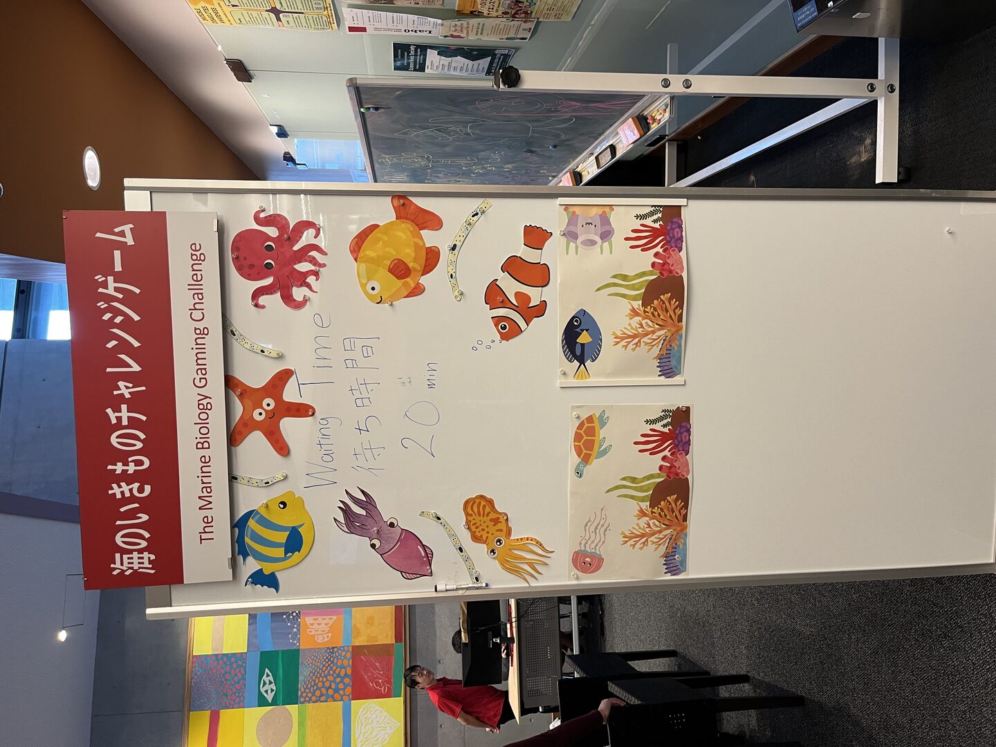
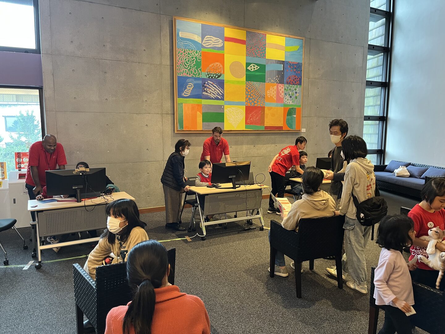
Later at OIST, I took this passion even further by setting up my own booth at the OIST Science Festival, where I introduced “The Marine Biology Computer Gaming Challenge.” Hundreds of kids gathered around, laughing, learning, and competing in the challenge. Seeing their faces light up as they explored science through gaming was truly rewarding. Many thanks to all the volunteers and administrators who helped me making this big event a success.
https://www.linkedin.com/posts/lajbner
Research Highlights
