

OCNC2006
Top | Schedule | Lectures | Posters | Projects | People
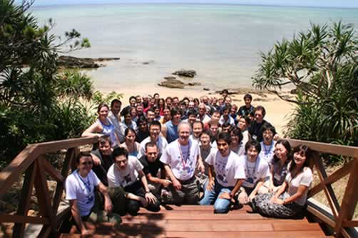
Okinawa Computational Neuroscience Course 2006
The goal of Okinawa Computational Neuroscience Course is to provide opportunities for young researchers with theoretical backgrounds to learn up-to-date neurobiological findings, and those with experimental backgrounds to have hands-on experience in computational modeling.
The special topic for this year's course is "Computing Neurons - What neurons compute; How we know by computing -"
We invite graduate students and postgraduate researchers to participate. The course will be held from June 26th through July 7th at an oceanfront Seminar House of Okinawa Institute of Science and Technology.
Okinawa Computational Neuroscience Course (OCNC 2006)
Date: June 26th to July 7th, 2006
Place: OIST Seaside House, Onna Village, Okinawa, Japan
Sponsors:
- Okinawa Institute of Science and Technology
- Nara Institute of Science and Technology
- Japanese Neural Network Society
Co-organizers:
- Upinder Bhalla, National Center for Biological Sciences, India
- Kenji Doya, Okinawa Institute of Science and Technology
- Shinya Kuroda, University of Tokyo
- Nicolas Le Novere, European Bioinformatics Institute
Advisory Board:
- Sydney Brenner, Okinawa Institute of Science and Technology
- Hiroaki Kitano, SONY Computer Science Laboratory
- Terrence Sejnowski, Salk Institute
- Susumu Tonegawa, MIT
Theme: Computing Neurons - What neurons compute; How we know by computing -
Lecturers:
- Upinder Bhalla, National Center for Biological Sciences
- Haruhiko Bito, University of Tokyo
- Sydney Brenner, Okinawa Institute of Science and Technology
- Yang Dan, University of California, Berkeley
- Erik De Schutter, University of Antwerp
- Kenji Doya, Okinawa Institute of Science and Technology
- Bard Ermentrout, University of Pittsburgh
- Geoffrey Goodhill, University of Queensland
- David Holcman, Weizmann Institute of Science
- Shin Ishii, Nara Institute of Science and Technology
- Shinya Kuroda, University of Tokyo
- Nicolas Le Novere, European Bioinformatics Institute
- Ion Moraru, University of Connecticut
- Felix Schuermann, Brain Mind Institute, EPFL
- Terrence Sejnowski, Salk Institute
- Susumu Tonegawa, MIT
- Jeff Wickens, University of Otago
Our brain is a network of billions of neurons, but even a single neuron is a fantastically complex computing device. Technology has made it possible to look into the detailed structure of dendritic branches, variety of ionic channels and receptors, molecular reactions at the synapses, and the network of genes that regulate all these. The challenge is to understand the meaning and function of these components of the neural machine. To do this we need to put together data from many experiments at different levels into a computational model, and to analyze the kinds of computation that single neurons and their networks can perform. This course invites graduate students and postgraduate researchers who are interested in studies integrating experimental and computational approaches for understanding cellular mechanisms of neurons.
Top | Schedule | Lectures | Posters | Projects | People
Schedule
| Sunday, June 25th | Check-in | |
| Monday, June 26th | ||
|
9:00-12:00
|
Susumu Tonegawa | |
|
14:00-15:00
|
Kenji Doya (Introduction) | |
| 15:00-17:00 | Guidance for Student Project | |
|
18:00-20:00
|
Welcome Party | |
| Tuesday, June 27th | ||
|
9:00-12:00
|
Geoffrey Goodhill | |
|
14:00-17:00
|
Jeff Wickens | |
| 17:00-18:00 | Poster presentation: 1 | |
| Wednesday, June 28th | ||
|
9:00-12:00
|
Bard Ermentrout | |
|
14:00-17:00
|
Terrence Sejnowski | |
| 17:00-18:00 | Poster presentation: 2 | |
| Thursday, June 29th | ||
|
9:00-12:00
|
Upinder Bhalla | |
|
14:00-17:00
|
Nicolas Le Novere | |
| 17:00-18:00 | Poster presentation: 3 | |
| Friday, June 30th | ||
|
9:00-12:00
|
Shinya Kuroda | |
|
13:30-18:00
|
OIST Initial Research Lab Tour | |
| Saturday, July 1st | ||
|
9:00-12:00
|
Erik De Schutter | |
|
14:00-17:00
|
Nicolas Le Novere | |
|
Sunday, July 2nd
|
||
| Day-off | ||
| Monday, July 3rd | ||
|
9:00-12:00
|
Sydney Brenner | |
|
14:00-17:00
|
Shin Ishii | |
| Tuesday, July 4th | ||
|
9:00-12:00
|
Felix Schuermann | |
|
14:00-17:00
|
Ion Moraru | |
| Wednesday, July 5th | ||
|
9:00-12:00
|
David Holcman | |
|
14:00-17:00
|
Haruhiko Bito | |
| Thursday, July 6th | ||
|
9:00-12:00
|
Yang Dan | |
|
14:00-17:00
|
Free discussion | |
| Friday, July 7th | ||
|
9:00-17:00
|
Presentations of Student projects | |
|
18:00-20:00
|
Farewell Party | |
| Saturday, July 8th | Check-out | |
Top | Schedule | Lectures | Posters | Projects | People
Lectures
Schedule
|
26 (mon)
|
27 (tue)
|
28 (wed)
|
29 (thu)
|
|
|---|---|---|---|---|
| Morning | ||||
| Afternoon |
Doya
|
|||
| 30 (fri) | 1 (sat) | 2 (sun) | 3 (mon) | |
| Morning |
Day-off
|
|||
| Afternoon |
Lab tour
|
|||
| 4 (tue) | 5 (wed) | 6 (thu) | 7 (fri) | |
| Morning |
Presentation of Student project
|
|||
| Afternoon |
Free discussion
|
|||
Lecture Title, Abstract, and Suggested Readings:
Susumu Tonegawa - "Molecular and circuit mechanisms for hippocampal memory studied by genetically engineered mice"
We study molecular, cellular, and neuronal circuit mechanisms underlying acquisition, consolidation and retrieval of hippocampus-dependent memory in rodents. Our primary approach is to generate cell type and adult-restricted knockout mice and characterize them using multifaceted methods including molecular and cellular biology, in vitro and in vivo electrophysiology, confocal and two photon microscopy and behavioral tasks. The data obtained to date indicate that NMDA receptor-mediated synaptic plasticity in area CA1 plays a pivotal role in spatial and other hippocampus dependent learning and memory. The same receptors and synaptic plasticity in area CA3 are dispensable for the acquisition of reference memory, but play an important role in “pattern completion”-based memory recall as well as in rapid, one trial-based learning. NMDA receptor function in dentate gyrus (DG) is also dispensable for reference memory, but is important in “pattern separation.” These studies attest the power of this multi-faceted approach in identifying mechanisms underlying cognition.
Suggested Readings:
Kenji Doya - "Computing Neurons: An Introduction"
Our brain is a network of billions of neurons, but even a single neuron is a fantastically complex computing device. Technology has made it possible to look into the detailed structure of dendritic branches, variety of ionic channels and receptors, molecular reactions at the synapses, and the network of genes that regulate all these. The challenge is to understand the meaning and function of these complex machinery. To do this we need to put together data from many experiments at different levels into a computational model, and to analyze the kinds of computation that single neurons and their networks can perform.
This introductory lecture aims to bring students from different disciplines to the common start line and to give overview of the forthcoming corse lectures. The topics will include the features of a neuron at different spatial and temporal scales, and computational and mathematical tools for reconstructing and analyzing them.
Suggested Readings:
Churchland PS, Sejnowski TJ (1992) The Computational Brain. MIT Press.
Koch C, Segev I (1998) Methods in Neuronal Modeling: From Ions to Networks, 2nd edition. MIT Press.
De Schutter E (2000) Computational Neuroscience: Realistic Modeling for Experimentalists. CRC press.
Fall CP, Marland ES, Wagner JM, Tyson JJ (2002) Computational Cell Biology. Springer Verlag.
Lecture slides (ppt)
Lecture movie (119.4 MB)
Geoffrey Goodhill - "Axon guidance and map formation"
For brains to function correctly their neurons must be wired up correctly. There is not enough information available in DNA to specify each connection individually, therefore brains must exploit more general rules to solve this wiring problem. These rules include following molecular gradients to find the right general region to
connect to, and then refining these initially crude connection patterns by neural activity. I will review recent experimental and theoretical advances in our understanding of the development of wiring in the brain, focusing particularly on gradient detection and map formation in the visual system.
Suggested Readings:
Jeff Wickens - "Local circuits and synaptic plasticity in the striatum"
The striatum of the mammalian brain receives excitatory input from almost every cortical area. It also receives a major dopaminergic input from the midbrain. Both types of input converge on the principal neurons of the striatum, which are spiny projection neurons. The spiny projection neurons are GABAergic and inhibit the output structures of the basal ganglia. The spiny projection neurons also make circuits with other striatal cells via their local collaterals. These anatomical features, together with other evidence, have inspired computational models of the striatum, including models of reinforcement-learning and winner-take-all inhibitory interactions. I shall sumarise the results of experimental work on synaptic plasticity in the corticostriatal pathway and on the nature of local circuit interactions among striatal neurons. These experimental results suggest important neural constraints on computational models of corticostriatal operations. In particular, lateral inhibition in the striatum is relatively sparse, unlike that assumed in winner-take-all network models. Also, dopamine-dependent plasticity of the cortical inputs seems to have strict temporal requirements that do not support the idea of an eligibility trace like that assumed in some reinforcement learning models. New models of the striatum and corticostriatal interactions are needed to understand the implications of these experimental findings for the information processing operations of the striatum.
Suggested Readings:
Czubayko U. and Plenz D. (2002). Fast synaptic transmission between striatal spiny projection neurons. Proc Natl Acad Sci U S A 99, 15746-9.
Koos T., Tepper J.M. and Wilson C.J. (2004). Comparison of IPSCs evoked by spiny and fast-spiking neurons in the neostriatum. J Neurosci, 24: 7916-22
Reynolds J., Hyland B.I. and Wickens J.R. (2001). A cellular mechanism of reward-related learning. Nature 413, 67-70.
Reynolds, J. N. and Wickens, J. R. (2002). Dopamine-dependent plasticity of corticostriatal synapses. Neural Netw 15, 507-21.
Tunstall M.J., Oorschot D.E., Kean A. and Wickens J.R. (2002). Inhibitory interactions between spiny projection neurons in the rat striatum. J Neurophysiol 88, 1263-1269.
Wickens J.R., Reynolds J.N.J. and Hyland B.I. (2003). Neural mechanisms of reward-related motor learning. Curr. Opin. Neurobiol. 13, 685-90.
Wickens J.R. (1997). Basal ganglia: Structure and computations. Network: Comp Neur Syst 8, 77-109.
Bard Ermentrout - "Neural oscillations"
Neural oscillations play roles in many moror patterns as well as possible roles in cognition and sensory coding. In this lecture I will introduce the mechanisms for production of oscillations in neurons and networks. Then I will turn to the derivation and description of phase resetting curves which play a fundamental role in our understanding of network oscillations. I will turn to weak coupling and how to analyze spatial networks of coupled oscillators. I will conclude with some reduced neuroal models and wave propagation.
Suggested Readings:
1) Tutorials in Mathematical Biosciences I : Mathematical Neuroscience (Lecture Notes in Mathematics / Mathematical Biosciences, Springer 2005)
2) Methods in Neuronal Modeling - 2nd Edition: From Ions to Networks (Computational Neuroscience) Koch & Segev editors, MIT Press
3) Dynamics of Coupled Map Lattices and of Related Spatially Extended Systems (Lecture Notes in Physics) (JR Chazottes B. Fernandez, ed)
4) N. Kopell and G.B. Ermentrout. "Mechanisms of phaselocking and
frequency control in pairs of coupled neural oscillators", for Hand-
book on Dynamical Systems, vol. 2: Toward applications. Ed. B.
Fiedler, Elsevier, pp 3-5. (2002)
5) Strogatz, S. [2000] "From Kuramoto to Crawford:exploring the onset of synchronization in populations of coupled oscillators", Physica D, 143:1-20
Terrence Sejnowski - "Calcium signaling in dendritic spines"
The significance of dendritic spines has been hotly debated since the time of Cajal. Recent experimental data suggest that by providing chemical compartmentalization, spines may act as functionally autonomous computational units. In particular, the spine may use calcium-binding proteins to compute the relative timing of glutamate release and postsynaptic spikes, whose pairing can induce long-term potentiation and depression. MCell is a computer program that uses Monte Carlo techniques to study cell signaling and takes into account the number and locations of ion channels and receptors, the kinetics of ion channel activation and the entry of calcium ions, and the intracellular diffusion of calcium and binding to calmodulin and other binding proteins. The results of the models can be directly tested with calcium imaging studies.
Suggested Readings:
Coggan J.S., Bartol T. M. Jr., Esquenazi E. I., Stiles J. R., Lamont S., Martone M. E., Berg D. K., Ellisman M. H., Sejnowski T. J. (2005). Evidence for Ectopic Neurotransmission at a Neuronal Synapse. Science, 39, 446-451, 2005
Lecture slides (ppt)
Lecture movie (236.3 MB)
Upinder Bhalla - "Signaling computations at the synapse: modeling molecular computation"
Signaling computations at the synapse: modeling molecular computation. Synapses are extremely compact computational structures. Although their physiological effect is normally thought of in terms of conductance changes, by far their greatest computational power arises through biochemical and molecular signalling. I will review work on frequency decoding, amplitude selectivity and pattern tuning in the synapse. I will also consider spatial aspects of synaptic computation. Finally, I will assert that the formation and maintenance of the synapse itself is a computational operation. I aim to convey the idea that computation, physiology, structure and cell biology of the synapse are essentially the same complex of processes, seen from different viewpoints.
In the second half of the class, I will use these computational issues as a motivation for building models. I will discuss how one performs computation with molecules. I will review some of basic chemistry and use simple reactions to build computational elements. I will give students a glimpse of how one goes about extracting the required information from experiments.
Suggested Readings:
Bray, D. (2002). Protein molecules as computational elements in living cells. Nature, 376, 307 - 312.
Bhalla, U.S. and Iyengar, R. (1999). Emergent properties of networks of Biological Signaling Pathways. Science, 15;283(5400): 381-7.
Bhalla, U.S. (2000). Simulations of biochemical signaling. In: Computational Neuroscience: Realistic Modeling for Experimentalists.Ed. E. De Schutter. CRC press.
Nicolas Le Novere - "Bioinformatics resources and standards for modeling neuronal signalling"
The development of quantitative models in neurobiology can be a lenghty and tedious work. However, an optimal use of existing data resources can improve much the efficiency of the process and the accuracy of the models. First, I will discuss the mining of biological data resources for model building and annotation. What are the useful resources out there? How do-they compare in terms of coverage, accuracy and maintenance? What can-we reuse in our models etc.
In order to expand our modelling toolkit, we need to reuse the models with a variety of software. I will present the standard formats to encode kinetic models (NeuroML, CellML, SBML...). In particular, I will spend a while on SBML, teaching how to use it to encode complex models of neuronal signalling.
I will finish on the problematic of model exchange and integration, describing ontology, standards and model databases that can help us developing large-scale models.
Suggested Readings:
Goddard NH et al. (2001). Towards NeuroML: model description methods for collaborative modelling in neuroscience. Philos Trans R Soc Lond B Biol Sci. 2001 Aug 29;356(1412):1209-28.
Hucka M., et al. (2003). The Systems Biology Markup Language (SBML): A Medium for Representation and Exchange of Biochemical Network Models. Bioinformatics, 19: 524-531.
Lloyd CM, et al. (2004) CellML: its future, present and past. Prog Biophys Mol Biol. 85(2-3):433-50.
Le Novère N., et al. (2005) Minimum Information Requested In the Annotation of biochemical Models (MIRIAM). Nature Biotechnology, 23: 1509-1515.
Le Novère N., et al. (2006) BioModels Database: A Free, Centralized Database of Curated, Published, Quantitative Kinetic Models of Biochemical and Cellular Systems Nucleic Acids Res., 34: D689-D691.
http://www.w3.org/XML/
http://sbml.org/
http://www.cellml.org/
http://www.neuroml.org/
http://www.ebi.ac.uk/biomodels/
http://doqcs.ncbs.res.in/
http://senselab.med.yale.edu/senselab/modeldb/
Lecture slides (pdf)
Lecture movie (197.7 MB)
Shinya Kuroda - "Modeling of synaptic plasticity"
Intercellular communication in neural circuits occurs at synapses, where molecular signaling networks decode ionic and biochemical patterns involving time, space and cellular context. The functional consequences of these computations are synaptic plasticity, including LTP and LTD. Induction of LTP and LTD depend on the timing between pre- and postsynaptic spiking, known as spike-timing-dependent synaptic plasticity (STDP) (1). I will talk about our computational STDP model in pyramidal neurons of the neocortex, where STDP is mainly NMDAR-dependent. We predicted that a certain property of NMDARs serves as a spike-timing detector in STDP (Urakubo, H. et al, in preparation). We experimentally validated this property by experiments (2) (data provided by Robert C. Froemke and Yang Dan at UC Berkeley). We also found that such property can explain complex spike-timing dependent synaptic plasticity such as spike-triplets and quadruplets (3). I will also talk about cerebellar LTD, which is mainly mGluR-dependent. We found that induction mechanism of LTD via Ca2+ depends on the regenerative cycle of IP3-dependent Ca2+-induced Ca2+-release (4), which induces the persistent activation of MAP kinase-dependent positive feedback loop (5).
Suggested Readings:
1. Dan, Y. and Poo, M. M. (2004). Spike timing-dependent plasticity of neural circuits. Neuron. 44, 23-30.
2. Froemke, R. C., Poo, M. M. and Dan, Y. (2005). Spike-timing-dependent synaptic plasticity depends on dendritic location. Nature. 434, 221-225.
3. Froemke, R. C. and Dan, Y. (2002). Spike timing-dependent synaptic modification induced by natural spike trains. Nature. 416, 433-438.
4. Doi T, Kuroda S, Michikawa T and Kawato M. (2005). Inositol 1,4,5-Trisphosphate-Dependent Ca2+ Threshold Dynamics Detect Spike Timing in Cerebellar Purkinje Cells. J Neurosci. Jan 26; 25(4): 950-61.
5. Kuroda S., Schweighofer N. and Kawato M. (2001). Exploration of Signal Transduction Pathways in Cerebellar Long-Term Depression by Kinetic Simulation. J Neurosci. Aug 1;21(15):5693-702.
Erik De Schutter - "Complex models of the cerebellar Purkinje cell: from molecules to learning"
First compartmental modeling methods will be introduced. After a brief description of cable theory relevant issues in creating passive compartmental models will be discussed, like obtaining good morphology and when is a neuron truly passive. Next building models with active dendrites will be described, using the Purkinje cell model as an example. This section will be wrapped up with a presentation of recent work on automated parameter searches for complex models which led to interesting insights into the parameter space of the Purkinje cell model.
The second part of the talk will focus on combined modeling and experimental studies. The effect of spines on diffusion of signaling molecules inside Purkinje cell dendrites will be described. Trapping by spines makes this diffusion process anomalous, a property which can be exploited in the context of learning. At a larger scale the effect of synaptic plasticity on Purkinje cell spiking will be discussed. Here we found that the active properties of the dendrite modulate the synaptic properties, resulting in unexpected effects on the spiking output.
Suggested Readings:
De Schutter E. and Bower J.M. (1994). Simulated responses of cerebellar Purkinje cells are independent of the dendritic location of granule cell synaptic inputs. Proceedings of the National Academy of Sciences USA 91: 4736-4740.
Achard P. and De Schutter E. (2006). Complex parameter landscape for a complex neuron model. PLOS Computational Biology. (in press)
Santamaria F., Wils S., De Schutter E. and Augustine, G.J.(2006). Anomalous diffusion in Purkinje cell dendrites caused by dendritic spines. Neuron.
Nicolas Le Novere - "Single-particle based simulation: How to avoid combinatorial explosion and take into account diffusion and topology"
Suggested Readings:
Morton-Firth, C. J., & Bray, D. (1998) Predicting temporal fluctuations in an intracellular signalling pathway. Journal of Theoretical Biology 192:117-128.
Shimizu, T. S., Aksenov, S. V., & Bray, D. (2003) A spatially extended stochastic model of the bacterial chemotaxis signalling pathway. Journal of Molecular Biology 329:291-309.
Tolle D., Le Novère N. (2006). Particle-based Stochastic Simulation in
Systems Biology. Current Bioinformatics, in the press. (preprint at:
http://www.ebi.ac.uk/compneur-srv/doc/Tolle2006.pdf)
Download the lecture slides (pdf) 1 2
Lecture movie (146.5 MB)
Sydney Brenner - "Computational aspects of neuron identity"
A central problem in the construction of nervous systems is the genetic representation of the "wiring diagram". The differences between the brains of mice and our brains must be specified by genes, but we know very little about how this is encoded and how the description is translated into the phenotype. In order to tackle this problem, it is necessary to have clear definition of cell type in the nervous system and we also need to reexamine the now murky notion of a gene. We will discuss these issues to provide a way to analyse the genetic basis of neuron identity.
Shin Ishii - "Bayesian reproduction of biological data"
In this lecture, I introduce Bayesian reverse modeling techniques applicable to various kinds of biological data; they are possibly spikes from a neural ensemble, behaviors from a human psychophysical experiment, gene expression time-series, or metabolite time-series observed in a metabolic pathway. If we can access a priori knowledge of the target biological system, Bayesian modeling provides us with a reliable tool for analyses by means of reverse engineering.
Suggested Readings:
Brown, E. N., Frank, L. M., Tang, D., Quirk, M. C., and Wilson, M. A. (1998). A statistical paradigm for neural spike train decoding applied to position prediction from ensemble firing patterns of rat hippocampal place cells. The Journal of Neuroscience, 18(18), 7411-7425.
Yoshida, W., and Ishii, S. (2006). Resolution of uncertainty in prefrontal cortex. Neuron, 50(5), 781-789.
Moles, C. G., Mendes, P., and Banga, J. R. (2003). Parameter estimation in biochemical pathways: A comparison of global optimization methods. Genome Research, 13, 2467-2474.
Crampin, E. J., Schnell, S., and McSharry P. E. (2004) Mathematical and computational techniques to deduce complex biochemical reaction mechanisms. Progress in Biophysics & Molecular Biology, 86, 77-112.
Felix Schuermann - "The Blue Brain Project"
The initial phase of the Blue Brain Project aims to reconstruct the detailed cellular structure and function of the neocortical column (NCC) of the young rat termed the Blue Column. This involves building 3D morphological and NEURON models of each type of neuron in the column, cloning a representative number of each neuron to capture the full morphological and electrical diversity, distributing neurons according to their correct proportions in each layer, interconnecting neurons according to their statistical connection rates as well as the pre and postsynaptic innervation patterns (numbers and distributions of synapses) and assigning functional synapse that capture the kinetics and dynamics of the different types of neocortical synapses. The Blue Column is being constructed in progressive versions where each progression reflects a greater level of biological accuracy. Version 1 was completed in December, 2005 which constituted 10¹000 morphologically complex neurons with minimal NEURON models and randomly interconnected. The latest version will be presented which includes the capture of the much of the morphological diversity, the different electrical classes of neurons modelled with combinations of detailed biologically accurate HH ion channels, different neurons distributed proportionately in different layers, neurons connected statistically according to biological probabilities, biologically accurate pre and postsynaptic synaptic innervations, synaptic models that capture the transmission rules for eocortical synapses.
Ion Moraru - "Spatial modeling using the Virtual Cell: a case study simulating calcium dynamics in Purkinje neurons dendritic spines"
Intracellular signaling regulates cellular behavior in ways more subtle and complex than once sought. The large number of interactions between participating molecules shows that there are no simple signaling "pathways", but rather a signaling "network". Often, compounding of many different rate laws creates nonlinear behavior. Additionally, specific cellular morphologies and subcellular microdomains can alter signal propagation and give rise to different local or global responses. As a result, it is difficult to properly understand these mechanisms by simple qualitative reasoning. Quantitative models and computer simulations are becoming increasingly important as an adjunct to experimental studies, but are traditionally quite difficult to create. During the first part of the lecture, we will review some of the challenges of computational cell biology, and I will introduce the Virtual Cell software environment, an unique model building and simulation framework targeted to biologists, that is particularly useful for 2D and 3D spatial modeling. The second part of the lecture will focus on theoretical studies of calcium signaling in dendrites of Purkinje neurons, where a variety of small differences in stimulation protocols can produce wildly different responses (e.g. no calcium transient, or calcium spikes confined to a single dendritic spine, or calcium waves along the dendritic shaft). We will show how building quantitative models and running simulations using the Virtual Cell helps to unravel this complexity. We will provide a possible explanation for the role of the apparent paradox between high density and low sensitivity of the IP3 receptors, putative mechanisms for coincidence detection and supralinearity, and analyze the role of spine morphology and membrane lipid diffusion in this system.
Suggested Readings:
The Virtual Cell website (http://vcell.org/): register for a free account, explore the software features, browse user documentation, tutorials, and published models.
Moraru I.I. and Loew L.M. (2005). Intracellular signaling: Spatial and Temporal Control. Physiology 20, 169-179.
Hernjak N., Slepchenko B.M., Fernald K., Fink C.C., Fortin D., Moraru I.I., Watras J. and Loew L.M. (2005). Modeling and analysis of calcium signaling events leading to long-term depression in cerebellar Purkinje cells. Biophys J. 89, 3790-3806.
David Holcman - Modeling receptor trafficking and synaptic activity"
The synaptic weight between a pre- and a postsynaptic neuron depends in part on the number of postsynaptic receptors. On the surface of neurons, receptors traffic by random motion in and out of a synaptic microstructure called the Postsynaptic Density (PSD). In the PSD, receptors can be stabilized at the membrane when they bind to scaffolding proteins.
In this talk, we will review first the modeling of brownian receptors, the theory of chemical reactions which involves few number and then we present recent mathematical computations related to the mean time it takes for a receptor to escape a micro-domain through small holes. When binding occurs, we will discuss how to obtain an expression of the Dwell time, which is the time spent by a receptor before it exits the PSD. Finally, a Markovian approach is presented and used to estimate the mean number of bound receptors at the PSD.
Because dendritic spines, the locus of the post-synaptic excitatory
connections, are highly motile structures, we will further discuss the role of this fast motility, which can be induced by a change in the neuronal activity and can regulate the number of receptors and thus the synaptic weight.
Suggested Readings:
Modeling:
A Guide to First-Passage Processes, Table of Contents, List of Errata, Exercises, & Book Reviews S. Redner, (Cambridge University Press, New York, 2001).
Theory and application of stochastic differential equations / Zeev Schuss Imprint New York : Wiley, c1980
D. Holcman, Z. Schuss. 2004. Escape Through a Small Opening: Receptor Trafficking in a Synaptic Membrane, J. of Statistical Physics. 117, 5/6 975 - 1014.
Biology:
Bredt DS, Nicoll RA. Related Articles, Links [Abstract] AMPA receptor trafficking at excitatory synapses. Neuron. 2003 Oct 9;40(2):361-79. Review.
Malinow R. Related Articles, Links [Free Full Text] AMPA receptor trafficking and long-term potentiation. Philos Trans R Soc Lond B Biol Sci. 2003 Apr 29;358(1432):707-14. Review.
Nicoll RA, Tomita S, Bredt DS. Related Articles, Links [Abstract] Auxiliary subunits assist AMPA-type glutamate receptors. Science. 2006 Mar 3;311(5765):1253-6. Review.
Choquet D, Triller A. Related Articles, Links [No abstract] The role of receptor diffusion in the organization of the postsynaptic membrane.
Nat Rev Neurosci. 2003 Apr;4(4):251-65. Review.
Haruhiko Bito - "How do neurons determine the number of axonal and dendritic processes? A simple question with a complex answer"
Neurons grow two characteristic processes, axons and dendrites. The specification of these processes, their outgrowth and their precise arborization are absolute prerequisites for formation of appropriate connections between neurons. These steps, which constitute the basis for establishment and maturation of neural circuits, represent central questions in neuroscience for which the molecular mechanisms still remain largely unsolved. Rho-family GTPases have been suggested to function as molecular switches involved in neuritogenesis and morphological specification of central neurons. However, the underlying biochemical mechanisms upstream and downstream of Rho small GTPases have remained obscure.
A Rho-target, p160ROCK, plays a key role in the regulation of early axonogenesis in cultured cerebellar granule neurons. We demonstrated that p160ROCK inhibition triggered immediate outgrowth of membrane ruffles and filopodia, followed by the generation of initial growth cone-like membrane domains from which axonal processes arose. Conversely, overexpression of dominant active forms of Rho or ROCK prevented the initiation of axon outgrowth. Further analyses indicated that the ROCK regulation of axonal outgrowth was likely to be mediated by multiple downstream effectors including LIMK-1. Taken together, Rho/ROCK pathway may constitute a gate critical for the initiation of axon outgrowth and the control of growth cone dynamics.
The establishment of dendritic arbors, which is critical for proper integration of synaptic inputs, is believed to be determined by genetically encoded cell intrinsic signals, as well as environmental signals from neighboring cells. Recent works have shed light to the critical roles of Semaphorin 3A, neurotrophins, Notch1, Slit-1, cadherin and beta-catenin in triggering and mediating some of the critical signaling events regulating various stages of dendritogenesis in the developing cerebral cortex. In addition, a wealth of work has shown that the formation of dendritic trees is shaped by neural activity. For example, surgical and pharmacological attenuation of sensory inputs impaired dendritic development in the visual cortex as well as in the barrel cortex, while in contrast, enriched environment promoted dendritic branching growth.
Recently, we have dealt with the issue of how the small GTPase activity may be turned on and off as a function of neuronal activity. We will present evidence that lipid raft-delimited signaling plays an essential role in coupling neuronal activity with small GTPase-mediated actin cytoskeletal remodeling during dendritogenesis.
Suggested Readings:
Da Silva JS, Dotti CG. Breaking the neuronal sphere: regulation of the actin cytoskeleton in neuritogenesis. Nat Rev Neurosci. 3: 694-704, 2002.
Bito H. (2003). Dynamic control of neuronal morphogenesis by rho signaling. J Biochem (Tokyo). 134: 315-319.
Van Aelst L. and Cline H.T. (2004). Rho GTPases and activity-dependent dendrite development. Curr Opin Neurobiol. 14: 297-304.
Konur S. and Ghosh A. (2005). Calcium signaling and the control of dendritic development. Neuron. 46: 401-405.
Luo L. and O'Leary DD. (2005). Axon retraction and degeneration in development and disease. Annu Rev Neurosci. 28:127-156.
Yang Dan - "Plasticity in the visual cortex: from synapse to perception"
In spike timing-dependent plasticity (STDP), presynaptic spiking tens of milliseconds before postsynaptic spiking induces long-term potentiation, whereas spiking of the opposite order induces long-term depression. This form of synaptic plasticity has been observed at glutamatergic synapses in a variety of neural circuits, and theoretical studies have shown that this synaptic learning rule endows neural circuits with great computational capacity. In this lecture, I will discuss experimental studies of STDP over the past several years in both in vivo and in vitro systems.
Suggested Readings:
Dan, Y. and Poo, M. M. (2004). Spike timing-dependent plasticity of neural circuits. Neuron. 44, 23-30.
Top | Schedule | Lectures | Posters | Projects | People
Posters
Tuesday, June 27th:
- Armin Biess
- Robert Brown
- Emma Coutts
- Jonathan Howard
- Wendy Huang
- Kotaro Kimura
- Mini Kurian
- Urs Koster
- Thomas McHugh
- Filip Piekniewski
- Anita Schmid
- Mete Balci
Wednesday, June 28th:
- Brenda Bloodgood
- Ricardo Carnieri
- Justin Dauwels
- Jaime Heiss
- Sachiko Ishida
- Yukihiro Nakamura
- Mohsen Omrani
- Manfred Oswald
- Dhanya Parameshwaran
- Malte Rasch
- Bobby Rohrkemper
- John Steephen
- Melanie Stefan
Thursday, June 29th:
- Dominic Tolle
- Shesharao Wanjerkhede
- Philipp Wolfrum
- Takashi Nakano
- Ketan Bajaj
- Naoki Honda
- Sungho Hong
- Shyful Islam
- Brian Lundstrom
- Aviv Mezer
- Hideaki Ogasawara
- Wiebke Potjans
- Ben Torben-Nielsen
Top | Schedule | Lectures | Posters | Projects | People
Projects
Student Projects
Most of the evening hours will be spent for student projects. Below are the three groups and tutors in charge of them:
A) Introduction to neural/molecular simulator platforms
Tutors: Quan Zou, Hidetoshi Urakubo, Aaron Gruber
B) Model construction from data
Tutors: Tomokazu Doi, Michele Giugliano, Rajnish Ranjan, Duncan Mortimer
C) Model analysis
Tutors: Yuichi Sakumura, Junichiro Yoshimoto, Klaus Stiefel
Top | Schedule | Lectures | Posters | Projects | People
People
Co-organizers:
- Upinder Bhalla, National Center for Biological Sciences, India
- Kenji Doya, Okinawa Institute of Science and Technology
- Shinya Kuroda, University of Tokyo
- Nicolas Le NovereNicolas Le Novere, European Bioinformatics Institute
Lecturers:
- Upinder Bhalla, National Center for Biological Sciences, India
- Haruhiko Bito, University of Tokyo
- Sydney Brenner, Okinawa Institute of Science and Technology
- Yang Dan, University of California, Berkeley
- Erik De Schutter, University of Antwerp
- Kenji Doya, Okinawa Institute of Science and Technology
- Bard Ermentrout, University of Pittsburgh
- Geoffrey Goodhill, University of Queensland
- David Holcman, Weizmann Institute of Science
- Shin Ishii, Nara Institute of Science and Technology
- Shinya Kuroda, University of Tokyo
- Nicolas Le Novere, European Bioinformatics Institute
- Ion Moraru, University of Connecticut
- Felix Schuermann, Brain Mind Institute, EPFL
- Terrence SejnowskiTerrence Sejnowski, Salk Institute
- Susumu TonegawaSusumu Tonegawa, MIT
- Jeff Wickens, University of Otago
Tutors:
- Tomokazu Doi, Osaka Bioscience Institute
- Michele Giugliano, Brain Mind Institute, EPFL
- Aaron Gruber, University of Maryland Baltimore
- Duncan Mortimer, University of Queensland
- Rajnish Ranjan, Brain Mind Institute, EPFL
- Yuichi Sakumura, Nara Institute of Science and Technology
- Klaus Stiefel, Salk Institute
- Hidetoshi Urakubo, University of Tokyo
- Junichiro Yoshimoto, Okinawa Institute of Science and Technology
- Quan Zou, UNIC, CNRS
Students:
- Ketan Bajaj, Indian Institute of Technology - Delhi, India
- Mete Balci, Istanbul Technical University
- Armin Biess, Weizmann Institute of Science
- Brenda Bloodgood, Harvard Medical School
- Robert Brown, University of California, Los Angeles
- Ricardo Carnieri, Nagoya University
- Emma Coutts, Heriot Watt University
- Justin Dauwels, RIKEN Brain Science Institute
- Jaime Heiss, Weizmann Institute of Science
- Naoki Honda, Nara Institute of Science and Technology
- Sungho Hong, University of Washington
- Jonathan Howard, King's College London
- Wendy Huang, University of California, San Francisco
- Sachiko Ishida, University of Tokyo
- Mohammad Shyful Islam, Nara Institute of Science and Technology
- Kotaro Kimura, National Institute of Genetics, Japan
- Urs Köster, University of Helsinki
- Mini Kurian, Arizona State University
- Brian Lundstrom, University of Washington
- Thomas McHugh, MIT
- Aviv Mezer, Tel Aviv University
- Yukihiro Nakamura, University of Tokyo
- Takashi Nakano, Okinawa Institute of Science and Technology
- Hideaki Ogasawara, ATR Computational Neuroscience Labs
- Mohsen Omrani, Theoretical Physics and Mathematics, Tehran
- Manfred Oswald, University of Otago
- Dhanya Parameshwaran, National Centre for Biological Sciences, India
- Filip Piekniewski, Nicolaus Copernicus University
- Wiebke Potjans, BCCN, Freiburg
- Malte Rasch, Graz University of Technology
- Robert Rohrkemper Jr., Institute of Neuroinformatics, Zurich
- Anita Schmid, University of Osnabruck
- John Eric Steephen, Indian Institute of Technology - Bombay
- Melanie Stefan, European Bioinformatics Institute
- Dominic Tolle, European Bioinformatics Institute
- Benjamin Torben-Nielsen, University Maastricht
- Shesharao Wanjerkhede, University Of Hyderabad
- Philipp Wolfrum, Frankfurt Institute for Advanced Studies